Copyright
©The Author(s) 2017.
World J Gastroenterol. Aug 28, 2017; 23(32): 5904-5912
Published online Aug 28, 2017. doi: 10.3748/wjg.v23.i32.5904
Published online Aug 28, 2017. doi: 10.3748/wjg.v23.i32.5904
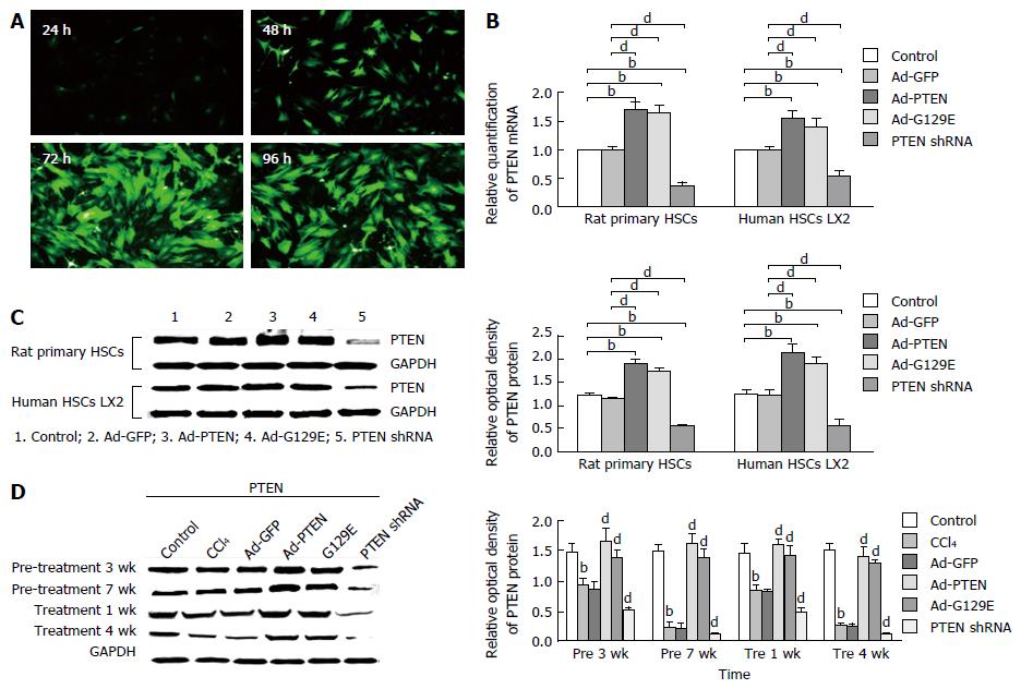
Figure 1 Effective modulation of the expression of phosphatase and tension homologue deleted on chromosome ten in vitro and in vivo.
A: Representative immunofluorescent image showing adenoviral transfection efficiency using Ad-GFP in rat primary HSCs; B: The mRNA expression of PTEN was quantitated for rat primary HSCs (left panel) and human LX-2 cells (right panel). Data are expressed as a relative value, and the error bars represent SE; C: Representative western blot showing the protein expression of PTEN (left panel), quantitated and expressed as relative optical density. The error bars represent SE. bP < 0.01 vs the control group; dP < 0.01 vs the Ad-GFP group; D: Representative western blot showing that the protein expression of PTEN in rat livers treated with Ad-PTEN was significantly enhanced in pre 1 wk, pre 3 wk, pre 5 wk, and pre 7 wk, compared with the Ad-GFP and control CCl4 groups. n = 3 for all experiments. GFP: Green fluorescent protein; HSCs: Hepatic stellate cells; PTEN: Phosphatase and tension homologue deleted on chromosome ten.
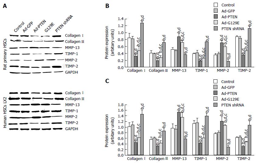
Figure 2 Phosphatase and tension homologue deleted on chromosome ten has a negative effect on collagen deposition in vitro.
A: Representative western blots of factors involved in the metabolism of fibrosis at 72 h posttransfection are shown in (A) and quantitated for rat primary HSCs in (B) and for human HSC LX2 cells in (C). Data in (B) and (C) are represented as mean ± SE. n = 3 for all groups. bP < 0.01 vs the control group; cP < 0.05 vs Ad-PTEN group; dP < 0.01 vs the Ad-GFP group. HSC: Hepatic stellate cell; PTEN: Phosphatase and tension homologue deleted on chromosome ten.
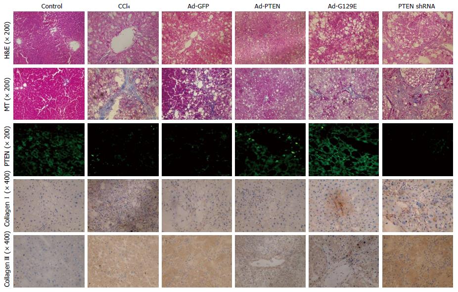
Figure 3 Phosphatase and tension homologue deleted on chromosome ten has protective effects on CCl4-induced rat hepatic fibrosis in vivo.
H&E stain (× 200) and MT stain (× 200) showed reduced hepatic cell necrosis and collagen deposition in liver tissue by over-expressed PTEN gene in Ad-PTEN and Ad-G129E groups. Immunofluorescent staining for PTEN (green) showed increased PTEN expression with Ad-PTEN and Ad-G129E recombinant adenovirus in both the prevention group or the treatment group. Immunohistochemical staining (bottom 2 rows) showed decreased collagen I and collagen III expression in hepatic tissues (× 400) with over-expression of PTEN induced by exogenous wild-type PTEN or G129E gene, whereas collagen I and collagen III expressions was reduced by PTEN shRNA. H&E: Hematoxylin and eosin; MT: Masson’s trichrome; PTEN: Phosphatase and tension homologue deleted on chromosome ten.
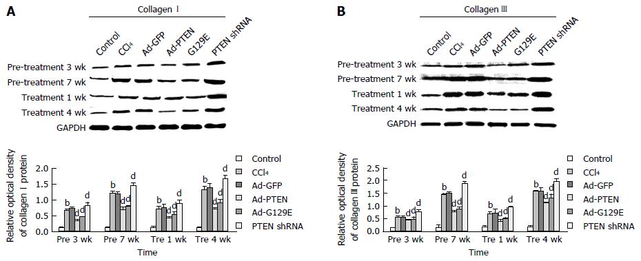
Figure 4 Phosphatase and tension homologue deleted on chromosome ten reduced collagen deposition in CCl4-induced fibrotic hepatic tissues.
Representative western blot of collagen I (A) and collagen III (B) expressions. Collagen I and collagen III expressions were quantitated in the bottom panels and represented as mean ± SE. bP < 0.01 vs the control group; dP < 0.01 vs the Ad-GFP group. PTEN: Phosphatase and tension homologue deleted on chromosome ten.
- Citation: Xie SR, An JY, Zheng LB, Huo XX, Guo J, Shih D, Zhang XL. Effects and mechanism of adenovirus-mediated phosphatase and tension homologue deleted on chromosome ten gene on collagen deposition in rat liver fibrosis. World J Gastroenterol 2017; 23(32): 5904-5912
- URL: https://www.wjgnet.com/1007-9327/full/v23/i32/5904.htm
- DOI: https://dx.doi.org/10.3748/wjg.v23.i32.5904


