Copyright
©The Author(s) 2017.
World J Gastroenterol. Jul 28, 2017; 23(28): 5253-5256
Published online Jul 28, 2017. doi: 10.3748/wjg.v23.i28.5253
Published online Jul 28, 2017. doi: 10.3748/wjg.v23.i28.5253
Figure 1 The radiographic image showed an esophago-bronchiole fistula from the anastomose to the right B1 bronchiole (arrow) and approximately 5 cm stenosis of the upper gastric tube (arrowhead).
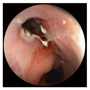
Figure 2 The epithelium of the esophago-bronchiole fistula was burned using argon plasma coagulation (arrow).
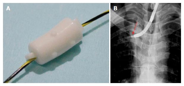
Figure 3 The endobronchial Watanabe spigot was penetrated through its long axis by the guidewire (A) (Push and Slide method[9]; B: Under fluoroscopic and endoscopic guidance, the endobronchial Watanabe spigot (arrow) was wedged into the esophago-bronchiole fistula.
Figure 4 The radiographic image just after the insertion showed the fistula occluded by the endobronchial Watanabe spigot (A, arrow); B: In the endoscopic image just after occlusion, it was confirmed that the endobronchial Watanabe spigot is in the target fistula.
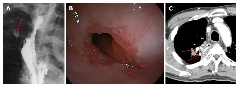
Figure 5 Since the endoscopic occlusion, the patient has not developed recurrence of the fistula for three years.
Two endobronchial Watanabe spigots remain (A, arrow); B: In the endoscopic image passed for three years after occlusion, the fistula has disappeared; C: In computed tomography passed for three years after occlusion, two endobronchial Watanabe spigots remain (arrow).
- Citation: Uesato M, Kono T, Akutsu Y, Murakami K, Kagaya A, Muto Y, Nakano A, Aikawa M, Tamachi T, Amagai H, Arasawa T, Muto Y, Matsubara H. Endoscopic occlusion with silicone spigots for the closure of refractory esophago-bronchiole fistula after esophagectomy. World J Gastroenterol 2017; 23(28): 5253-5256
- URL: https://www.wjgnet.com/1007-9327/full/v23/i28/5253.htm
- DOI: https://dx.doi.org/10.3748/wjg.v23.i28.5253


