Copyright
©The Author(s) 2017.
World J Gastroenterol. Jul 21, 2017; 23(27): 4867-4878
Published online Jul 21, 2017. doi: 10.3748/wjg.v23.i27.4867
Published online Jul 21, 2017. doi: 10.3748/wjg.v23.i27.4867
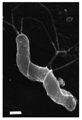
Figure 1 Morphology of Helicobacter pylori.
S-shaped H. pylori with five to seven sheathed polar flagella. Field emission SEM, bar = 0.5 μm. (Field emission SEMs courtesy of L. Thompson and negative stains courtesy of S. Danon, School of Microbiology and Immunology, University of New South Wales). From: Helicobacter pylori: Physiology and Genetics. Mobley HLT, Mendz GL, Hazell SL, editors. Washington (DC): ASM Press; 2001. Chapter 6, Morphology and Ultrastructure[54].
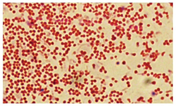
Figure 2 Microscopic images of coccoid forms of Helicobacter pylori after 6-d exposure to antibiotics.
From: Faghri et al[65].
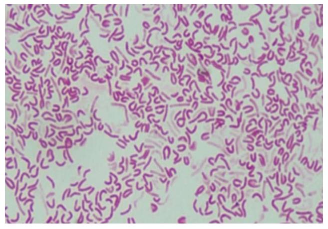
Figure 3 Microscopic images of morphological forms of Helicobacter pylori after exposure to antibiotics: S-shaped, U-shaped, C-shaped and coccoid-shaped.
From: Faghri et al[65].
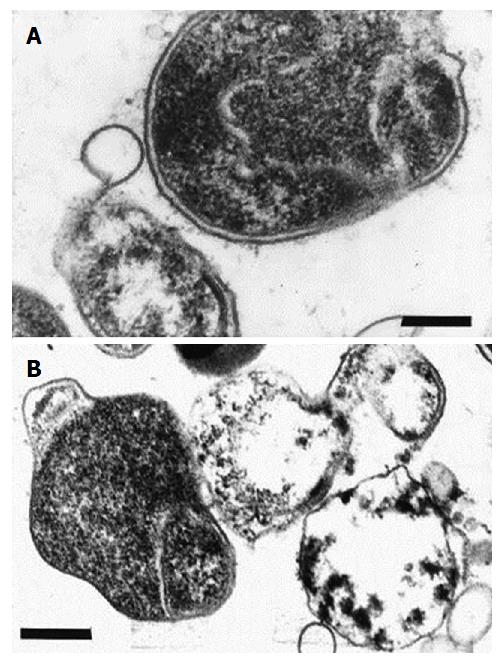
Figure 4 Electronograms of coccoid forms of Helicobacter pylori.
A: Initial ingrowth in the periplasmic space resulting in the formation of U-shaped cells; B: Conversion to the coccoid form. Ultrathin section, bar = 0.5 μm. From: Helicobacter pylori: Physiology and Genetics. Mobley HLT, Mendz GL, Hazell SL, editors. Washington (DC): ASM Press; 2001. Chapter 6, Morphology and Ultrastructure[54].
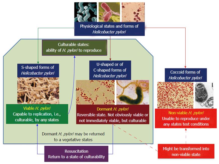
Figure 5 Major physiological states and forms of Helicobacter pylori[101].
H. pylori: Helicobacter pylori.
- Citation: Reshetnyak VI, Reshetnyak TM. Significance of dormant forms of Helicobacter pylori in ulcerogenesis. World J Gastroenterol 2017; 23(27): 4867-4878
- URL: https://www.wjgnet.com/1007-9327/full/v23/i27/4867.htm
- DOI: https://dx.doi.org/10.3748/wjg.v23.i27.4867


