Copyright
©The Author(s) 2017.
World J Gastroenterol. Jun 28, 2017; 23(24): 4467-4472
Published online Jun 28, 2017. doi: 10.3748/wjg.v23.i24.4467
Published online Jun 28, 2017. doi: 10.3748/wjg.v23.i24.4467
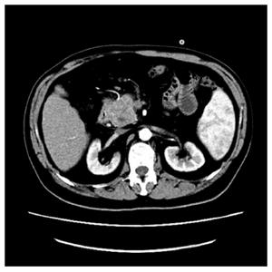
Figure 1 Abdominal computed tomography scan findings.
A 4.2 cm × 4.1 cm hypodense mass within the head of the pancreas.
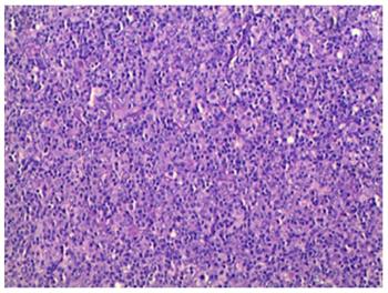
Figure 2 Histopathological examination.
Large atypical lymphoid cells were scattered among small reactive lymphocytes and less often histiocytes (HE, × 20).
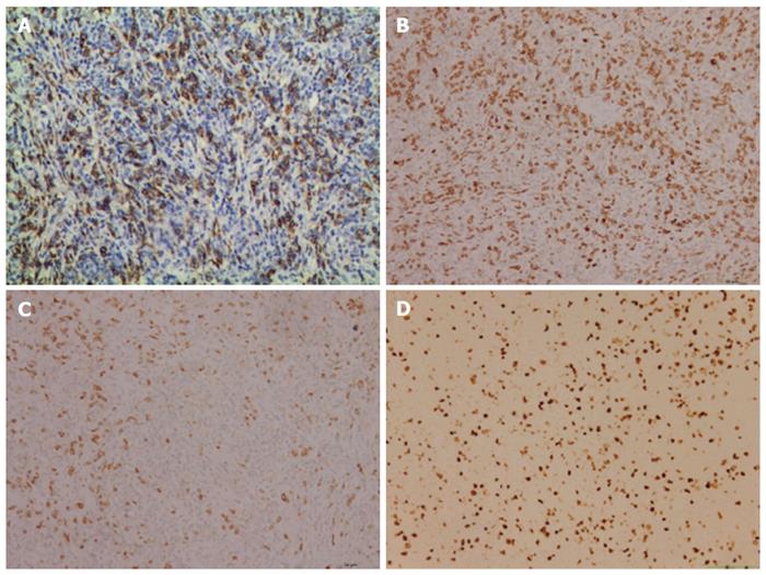
Figure 3 Immunohistochemistry studies.
A: The dispersed CD20 positive large B cells (× 20); B: Numerous CD3 positive small T cells in the background (× 20); C: CD68 positive histiocytes in the background (× 20); D: Ki-67 expression of neoplastic cells (× 20).
- Citation: Zheng SM, Zhou DJ, Chen YH, Jiang R, Wang YX, Zhang Y, Xue HL, Wang HQ, Mou D, Zeng WZ. Pancreatic T/histiocyte-rich large B-cell lymphoma: A case report and review of literature. World J Gastroenterol 2017; 23(24): 4467-4472
- URL: https://www.wjgnet.com/1007-9327/full/v23/i24/4467.htm
- DOI: https://dx.doi.org/10.3748/wjg.v23.i24.4467


