Copyright
©The Author(s) 2017.
World J Gastroenterol. Jun 28, 2017; 23(24): 4369-4380
Published online Jun 28, 2017. doi: 10.3748/wjg.v23.i24.4369
Published online Jun 28, 2017. doi: 10.3748/wjg.v23.i24.4369
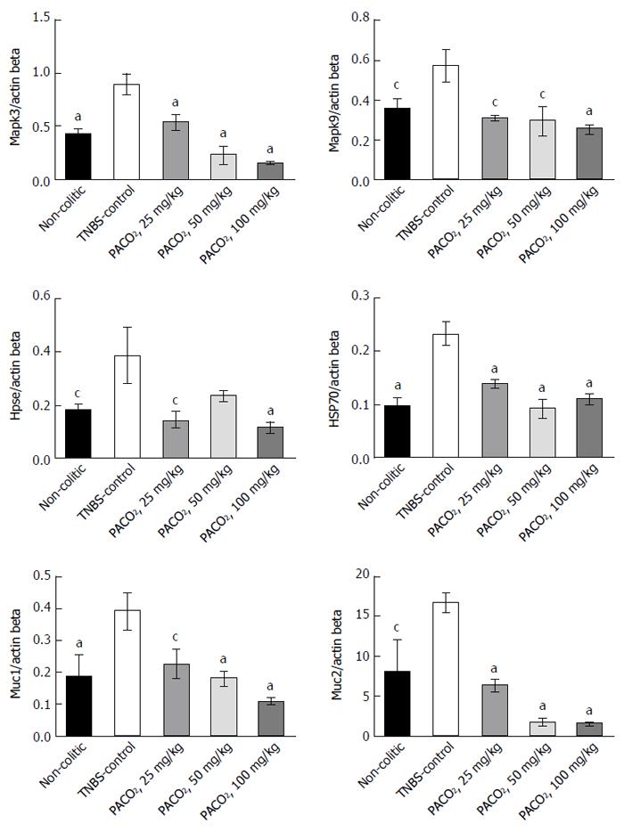
Figure 1 Effects of the PACO2 standardized extract (25, 50 or 100 mg/kg) in the gene expression of Mapk3, Mapk9, heparanase, Hsp70, Muc1 and Muc2 in acute phase response of intestinal inflammation induced by trinitrobenzenesulphonic acid.
Data are expressed as mean ± standard error of the mean. cP ≤ 0.05, aP ≤ 0.01 vs TNBS control group.
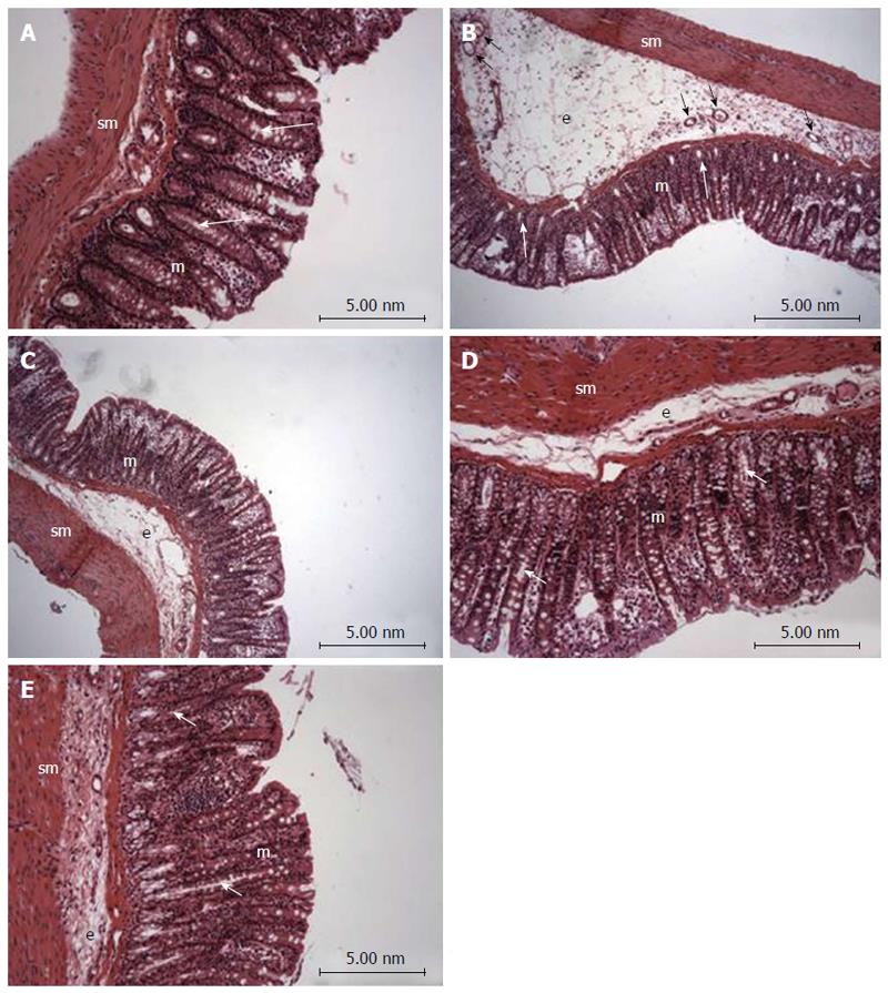
Figure 2 Photomicrography (hematoxylin and eosin) of colon samples from different experimental groups.
Mucosa (m), tubular glands (white arrows), blood vessels (black arrow), submucosa (sm) and edema (e). In A (non-colitic group): the mucosa (m) contains numerous straight tubular glands (white arrows) with many lightly stained goblet cells; the crypt (black arrow), submucosa (sm) and luminal epithelium was intact with a typical morphology; In B (TNBS control group): tubular glands (white arrows) were reduced and the goblet cells were atypical; a disruption with extensive edema (e) and blood vessels (black arrow) can be observed. In PACO2-treated groups, colon cytoarchitecture was recovering and included the restoration of tubular glands (white arrows) containing goblet cells, especially at the doses of 50 mg/kg (D) and 100 mg/kg (E); edema (e) was significantly reduced in animals treated with at doses of 50 mg/kg (D) and 100 mg/kg (E) and in a lower proportion in animals treated with doses of 25 mg/kg (C), when compared to the TNBS control group (B).
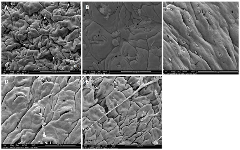
Figure 3 Electromicrographs (scanning electron microscopy) of colon samples from different experimental groups.
Crypts (white arrows), goblet cells (black arrows) and mucin (m). In A (non-colitic group): regular mucosal architecture with polygonal units as structural subunits, regular microvilli giving a smooth velvety appearance, crypts (white arrows), goblet cells (black arrows) and mucin (m) extruded; In B (TNBS control group): complete loss of the smooth velvety appearance and polygonal shape of absorptive cells, hyperplasia of goblet cells (white arrows), absence of mucin extrusion and crypts, characteristics of ulcerative colitis and Crohn’s disease; In C (PACO2 25 mg/kg-treated group): slight recovery of polygonal shape (black arrows) of absorptive cells, however the number of goblet cells remained elevated; In D (PACO2 50 mg/kg-treated group): smaller number of goblet cells, greater recovery of polygonal appearance (black arrows) and increased mucin (m) compared to TNBS control group; In E (PACO2 100mg/kg-treated group): good recovery of mucosa architecture, with goblet cells (black arrows) similar to healthy animals including crypts (white arrows) and mucin (m) for protection.
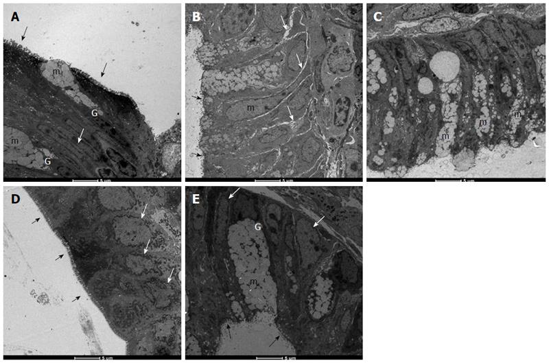
Figure 4 Electromicrographs (transmission electron microscopy) of colon samples from different experimental groups.
Goblet cells (G), mucus granules (m); Microvilli (black arrows); nucleus in a basal position (white arrow); edema (grey arrows). In A (non-colitic group): colonic goblet cells (G) showing the mucus granules (m); absorptive epithelial cells with a well-developed brush border containing numerous regular microvilli (black arrows) typical of enterocytes and its nucleus in a basal position (n) shows chromatin (white arrow); In B (TNBS control group): mucin granules in colonic goblet cells are reduced and disarranged (m); surface cells have no distinct brush border with atypical microvilli (black arrow); the intercellular space was enhanced as a signal of edema (white arrows); In C (PACO2 25 mg/kg-treated group): mucin granules in colonic goblet cells are reduced and disarranged (m), but intercellular edema is reduced (white arrows); In D (PACO2 50 mg/kg-treated group): juxtaposed colonocytes with nucleus in basal position (white arrows) and preserved microvilli (black arrows); In E (PACO2 100 mg/kg-treated group): goblet cells (G) showing the mucus granules (m) similar to healthy animals; nucleus in basal position (yellow arrows); mild intercellular edema and atypical microvilli (black arrows).
- Citation: Almeida Junior LD, Quaglio AEV, de Almeida Costa CAR, Di Stasi LC. Intestinal anti-inflammatory activity of Ground Cherry (Physalis angulata L.) standardized CO2 phytopharmaceutical preparation. World J Gastroenterol 2017; 23(24): 4369-4380
- URL: https://www.wjgnet.com/1007-9327/full/v23/i24/4369.htm
- DOI: https://dx.doi.org/10.3748/wjg.v23.i24.4369


