Copyright
©The Author(s) 2017.
World J Gastroenterol. Jun 28, 2017; 23(24): 4324-4329
Published online Jun 28, 2017. doi: 10.3748/wjg.v23.i24.4324
Published online Jun 28, 2017. doi: 10.3748/wjg.v23.i24.4324
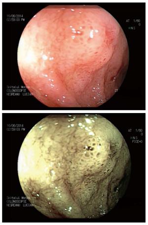
Figure 1 Crohn’s disease patient WL and flexible spectral imaging color enhancement.
Better delineation of ileal edema and ulceration using FICE.
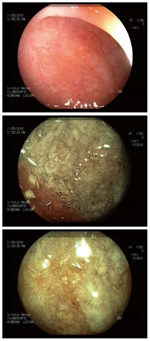
Figure 2 Ulcerative colitis-subtle lesions indicating recurrence getting more evident with flexible spectral imaging color enhancement.
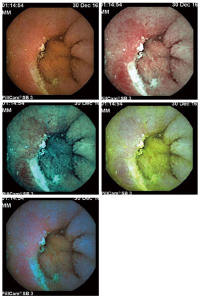
Figure 3 Capsule images of jejunal ulceration in WL flexible spectral imaging color enhancement 1, 2, 3 and blue mode.
- Citation: Goran L, Negreanu L, Negreanu AM. Role of new endoscopic techniques in inflammatory bowel disease management: Has the change come? World J Gastroenterol 2017; 23(24): 4324-4329
- URL: https://www.wjgnet.com/1007-9327/full/v23/i24/4324.htm
- DOI: https://dx.doi.org/10.3748/wjg.v23.i24.4324


