Copyright
©The Author(s) 2017.
World J Gastroenterol. Jun 7, 2017; 23(21): 3900-3906
Published online Jun 7, 2017. doi: 10.3748/wjg.v23.i21.3900
Published online Jun 7, 2017. doi: 10.3748/wjg.v23.i21.3900
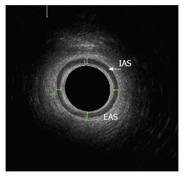
Figure 1 Internal anal sphincter thickness is defined as the mean thickness of the internal anal sphincter at 12, 3, 6, 9 o’clock positions in the mid-axial plane.
IAS: Internal anal sphincter; EAS: External anal sphincter.
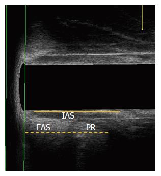
Figure 2 The Internal anal sphincter length (solid line) and the length of external anal sphincter plus puborectalis muscle (dotted line) were measured in the mid-sagittal plane.
IAS: Internal anal sphincter; EAS: External anal sphincter; PR: Puborectalis muscle.
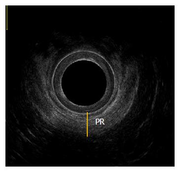
Figure 3 Thickness of puborectalis muscle was measured in the distal-axial plane (solid line).
PR: Puborectalis muscle.
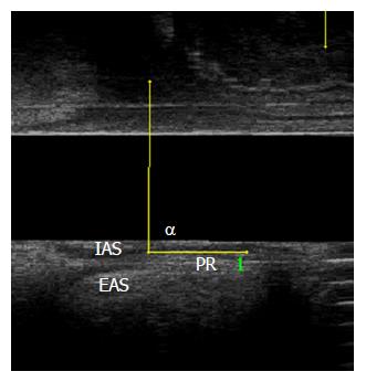
Figure 4 Puborectalis angle (α angle).
IAS: Internal anal sphincter; EAS: External anal sphincter; PR: Puborectalis muscle.
Figure 5 Puborectalis angle during resting phase (A) and straining phase (B) in chronic proctalgia and normal control groups (P < 0.
05). CP: Chronic proctalgia.
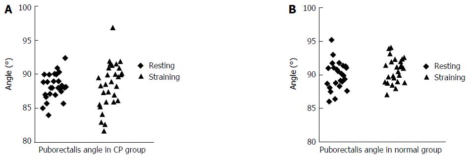
Figure 6 Puborectalis angle during resting and straining phases in the chronic proctalgia group (A) (P > 0.
05) and normal control group (B) (P < 0.05). CP: Chronic proctalgia.
- Citation: Xue YH, Ding SQ, Ding YJ, Pan LQ. Role of three-dimensional endoanal ultrasound in assessing the anal sphincter morphology of female patients with chronic proctalgia. World J Gastroenterol 2017; 23(21): 3900-3906
- URL: https://www.wjgnet.com/1007-9327/full/v23/i21/3900.htm
- DOI: https://dx.doi.org/10.3748/wjg.v23.i21.3900


