Copyright
©The Author(s) 2017.
World J Gastroenterol. Jan 14, 2017; 23(2): 366-372
Published online Jan 14, 2017. doi: 10.3748/wjg.v23.i2.366
Published online Jan 14, 2017. doi: 10.3748/wjg.v23.i2.366
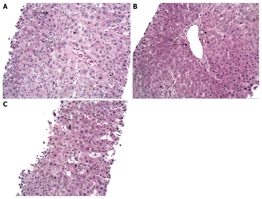
Figure 1 Liver biopsy.
A: The lobular parenchyma has marked cholestasis (arrow) with a zone 3 accentuation, associated with occasional feathery hepatocyte degeneration (arrowhead) and mild inflammation; B: Portal tract with portal vein (arrow) and two branches of hepatic arterioles (arrowheads) with missing bile duct; C: Ito cell lipidosis (arrowheads) were also seen. Hematoxylin-eosin staining, magnification × 200.
- Citation: Bakhit M, McCarty TR, Park S, Njei B, Cho M, Karagozian R, Liapakis A. Vanishing bile duct syndrome in Hodgkin’s lymphoma: A case report and literature review. World J Gastroenterol 2017; 23(2): 366-372
- URL: https://www.wjgnet.com/1007-9327/full/v23/i2/366.htm
- DOI: https://dx.doi.org/10.3748/wjg.v23.i2.366


