Copyright
©The Author(s) 2017.
World J Gastroenterol. Jan 14, 2017; 23(2): 256-264
Published online Jan 14, 2017. doi: 10.3748/wjg.v23.i2.256
Published online Jan 14, 2017. doi: 10.3748/wjg.v23.i2.256
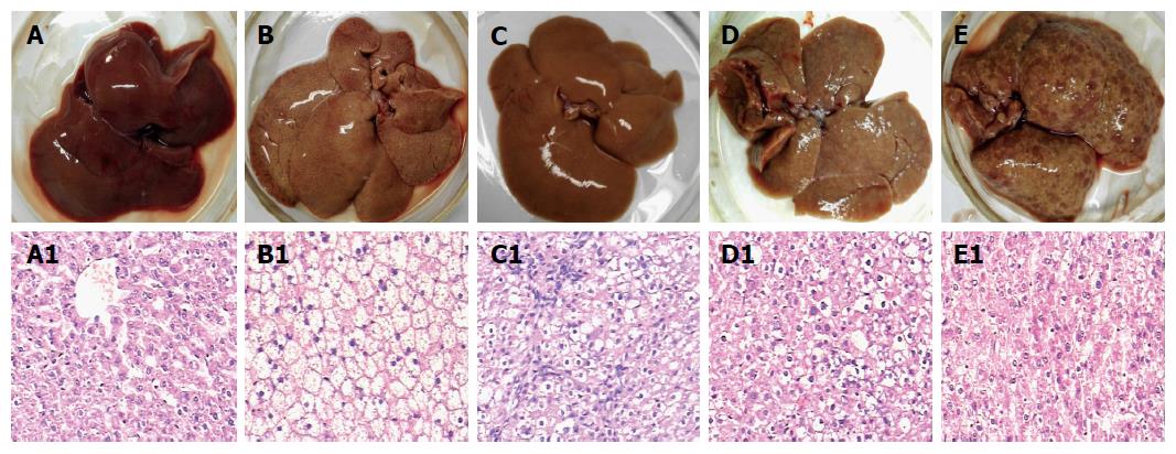
Figure 1 Rat liver tissues and their pathological examination.
Liver alterations after the rats were sacrificed at different stages during hepatocyte malignant transformation. A: A representative specimen from the control rats with normal diet; B: A representative specimen from the rats with high-fat diet without 2-fluorenyl acetamide (2-FAA); C: A representative specimen from the rats with high-fat diet containing 2-FAA, at early stage; D: A representative specimen from the rats with high-fat diet containing 2-FAA, at interim stage; and E: A representative specimen from the rats with high-fat diet containing 2-FAA, at later stage. The liver sections were examined with hematoxylin and eosin staining and then divided into the control (A1), fatty liver (B1), degeneration (C1), precancerous (D1), and cancerous (E1) groups; A1-E1: The original magnification of the corresponding rat liver sections was × 200.
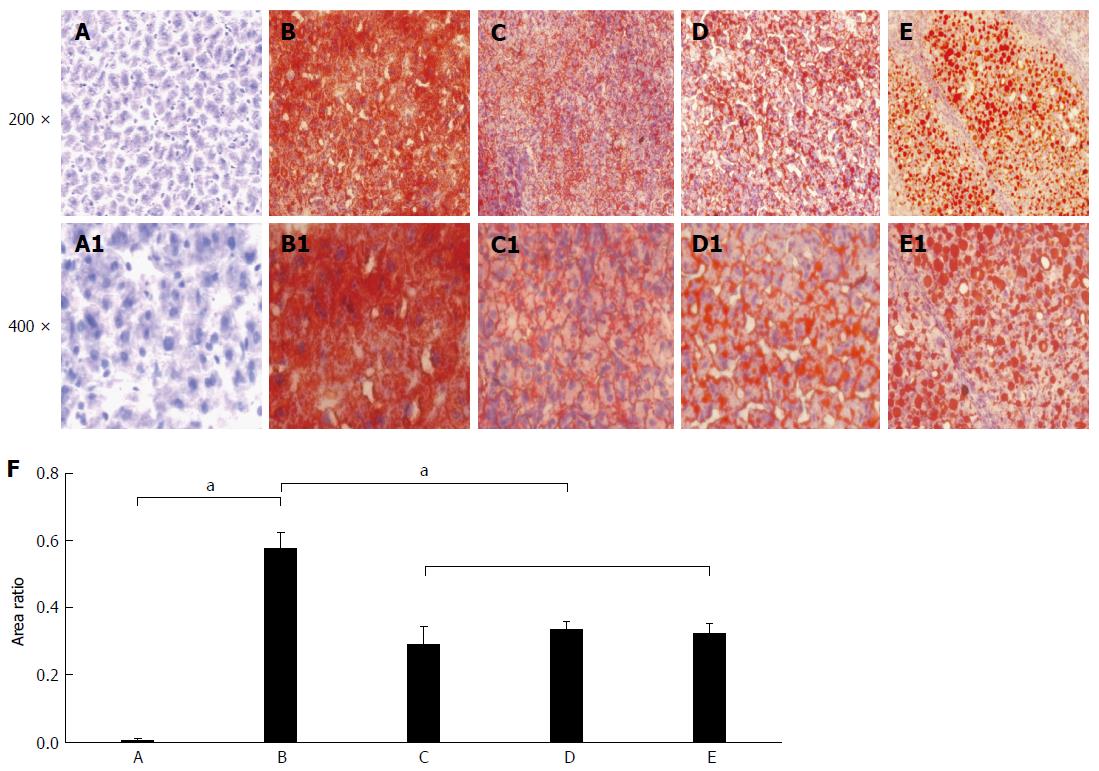
Figure 2 Rat hepatic lipid accumulation in the different groups after liver section staining with the Oil Red O.
The status of hepatic lipid accumulation in the different rat groups was analyzed after liver section staining with the Oil Red O method. A: Liver sections of control rats with normal diet; B: Liver sections of the fatty liver rats with high-fat diet without 2-fluorenylacetamide (2-FAA); C: Liver sections of the degeneration rats with high-fat diet containing 2-FAA, at early stage; D: Liver sections of precancerous rats with high-fat diet containing 2-FAA, at interim stage; E: Liver sections of cancerous rats with high-fat diet containing 2-FAA, at later stage; F: Relative quantitative analysis of fats in the liver tissue; A-E: The original magnification was × 200; A1-E1: The original magnification was × 400. aP < 0.05.
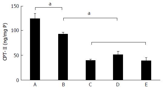
Figure 3 Dynamic alteration of carnitine palmitoyl transferase-II expression in rat livers.
The CPT-II contents of rat hepatic tissues in the different rat groups were analyzed during hepatocyte malignant transformation. A: Liver sections of control rats with normal diet; B: Liver sections of the fatty liver rats with high-fat diet without 2-fluorenylacetamide (2-FAA); C: Liver sections of the degeneration rats with high-fat diet containing 2-FAA, at early stage; D: Liver sections of precancerous rats with high-fat diet containing 2-FAA, at interim stage; E: Liver sections of cancerous rats with high-fat diet containing 2-FAA, at later stage. aP < 0.05. CPT-II: Carnitine palmitoyl transferase-II.
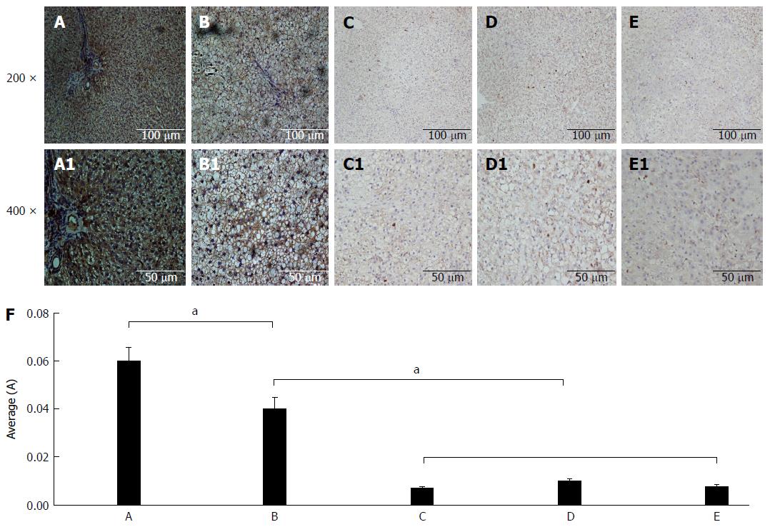
Figure 4 Immunohistochemical analysis of carnitine palmitoyl transferase II expression.
The CPT-II contents of rat hepatic tissues in the different rat groups were analyzed by immunohistochemistry. A: Liver sections of control rats with normal diet; B: Liver sections of the fatty liver rats with high-fat diet without 2-fluorenyl acetamide (2-FAA); C: Liver sections of the degeneration rats with high-fat diet containing 2-FAA, at early stage; D: Liver sections of precancerous rats with high-fat diet containing 2-FAA, at interim stage; E: Liver sections of cancerous rats with high-fat diet containing 2-FAA, at later stage; F: The relative quantitative analysis of CPT-II in the liver tissue; A-E: The original magnification was × 200; A1-E1: The original magnification was × 400. aP < 0.05. CPT-II: Carnitine palmitoyl transferase-II.
- Citation: Gu JJ, Yao M, Yang J, Cai Y, Zheng WJ, Wang L, Yao DB, Yao DF. Mitochondrial carnitine palmitoyl transferase-II inactivity aggravates lipid accumulation in rat hepatocarcinogenesis. World J Gastroenterol 2017; 23(2): 256-264
- URL: https://www.wjgnet.com/1007-9327/full/v23/i2/256.htm
- DOI: https://dx.doi.org/10.3748/wjg.v23.i2.256


