Copyright
©The Author(s) 2017.
World J Gastroenterol. May 21, 2017; 23(19): 3496-3504
Published online May 21, 2017. doi: 10.3748/wjg.v23.i19.3496
Published online May 21, 2017. doi: 10.3748/wjg.v23.i19.3496
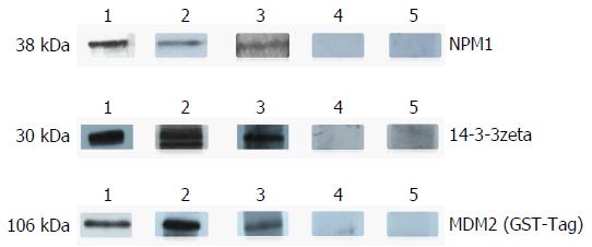
Figure 1 Western blot analysis of representative sera of three anti-tumor-associated antigens autoantibodies assessed by enzyme-linked immunosorbent assay.
Lane 1: The polyclonal anti-NPM1 autoantibody and anti-14-3-3zeta autoantibody were used as positive control; Lanes 2 and 3: Two representative AFP-negative HCC serum samples which were positive in ELISA also had strong reactivity to 14-3-3zeta recombinant protein in western blot analysis; Lanes 4 and 5: Randomly selected chronic liver disease sera and normal human control, respectively, with negative reactivity to 14-3-3zeta recombinant protein. AFP: Alpha fetoprotein; ELISA: Enzyme-linked immunosorbent assay; HCC: Hepatocellular carcinoma.
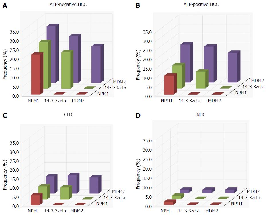
Figure 2 Analysis to determine the presence or absence of co-expression of antibodies to any combination of two of the three tumor-associated antigens in α fetoprotein-negative hepatocellular carcinoma, α fetoprotein-positive hepatocellular carcinoma, chronic liver disease and normal human control.
The height of the bar represents the percentage of sera with co-expression of two antibodies, e.g., NPM1 antibody with 14-3-3zeta antibody, and NPM1 antibody with MDM2 antibody. AFP: Alpha fetoprotein; CLD: Chronic liver disease; HCC: Hepatocellular carcinoma; NHC: Normal human control; TAAs: Tumor-associated antigens.
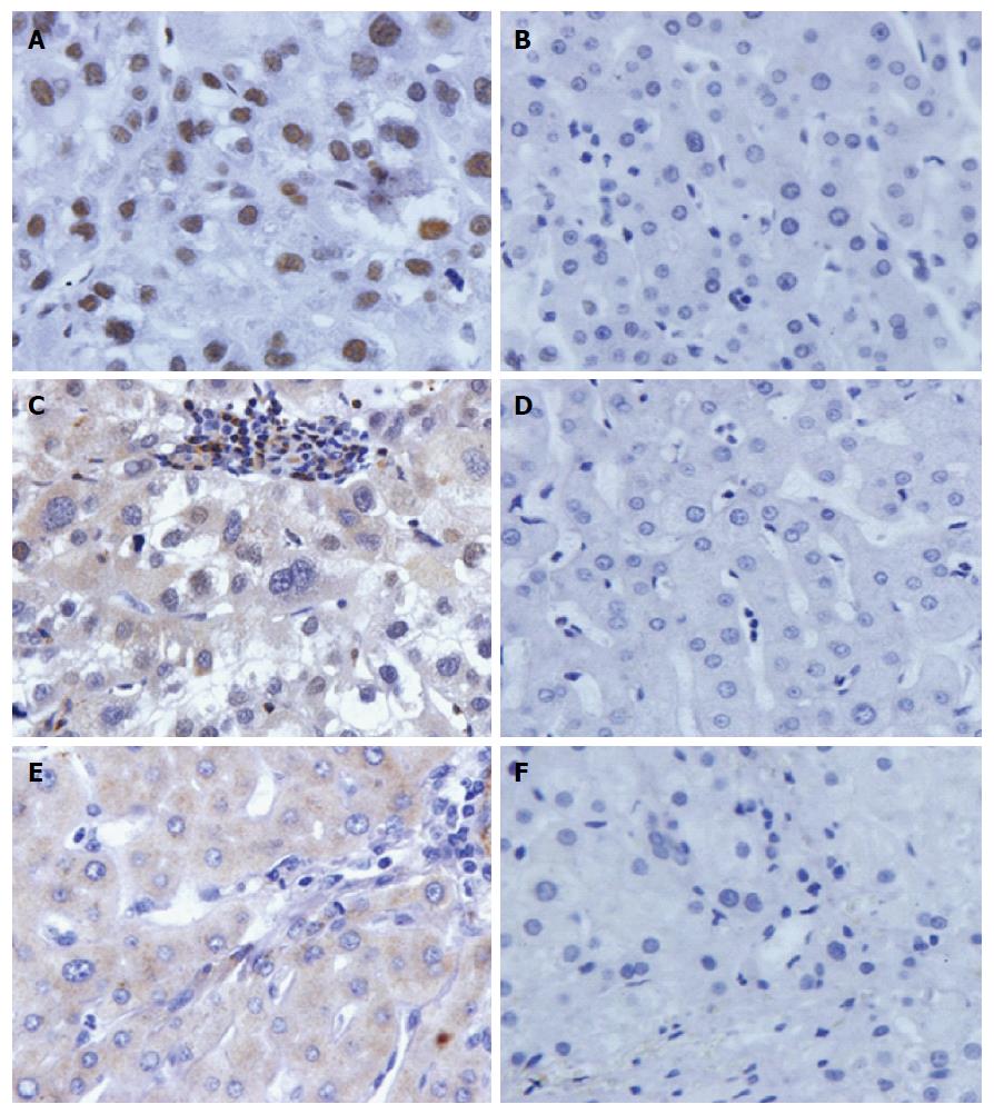
Figure 3 Expression of NPM1, 14-3-3zeta and MDM2 in α-fetoprotein-negative hepatocellular carcinoma tissues and normal hepatic tissues by immunohistochemistry.
The three polyclonal anti-TAAs antibodies were used as a primary antibody to detect their expression in liver cancer and normal hepatic tissues. A and B: HCC tissue with positive staining and normal hepatic tissue with negative staining in anti-NPM1 antibody; C and D: HCC tissue with strong positive staining and normal hepatic tissue with negative staining in anti-14-3-3zeta antibody; E and F: HCC tissue with strong positive staining and normal hepatic tissue with negative staining in anti-MDM2 antibody. AFP: Alpha fetoprotein; HCC: Hepatocellular carcinoma; TAAs: Tumor-associated antigens.
- Citation: Wang T, Liu M, Zheng SJ, Bian DD, Zhang JY, Yao J, Zheng QF, Shi AM, Li WH, Li L, Chen Y, Wang JH, Duan ZP, Dong L. Tumor-associated autoantibodies are useful biomarkers in immunodiagnosis of α-fetoprotein-negative hepatocellular carcinoma. World J Gastroenterol 2017; 23(19): 3496-3504
- URL: https://www.wjgnet.com/1007-9327/full/v23/i19/3496.htm
- DOI: https://dx.doi.org/10.3748/wjg.v23.i19.3496


