Copyright
©The Author(s) 2017.
World J Gastroenterol. Apr 28, 2017; 23(16): 2948-2956
Published online Apr 28, 2017. doi: 10.3748/wjg.v23.i16.2948
Published online Apr 28, 2017. doi: 10.3748/wjg.v23.i16.2948
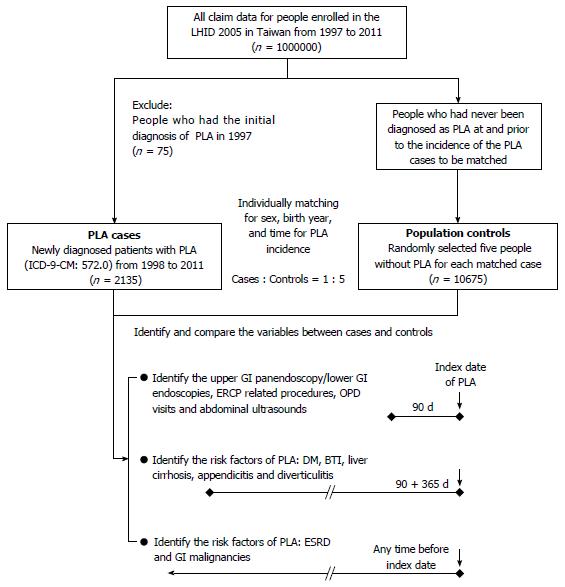
Figure 1 Flow chart for selecting cases and controls and identifying comorbidity and examinations.
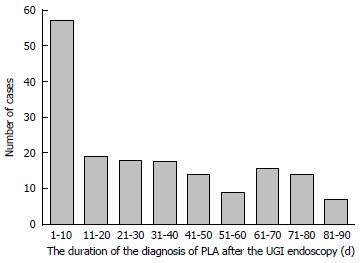
Figure 2 Frequency and duration of the diagnosis of pyogenic liver abscess after the first upper gastrointestinal panendoscopy examination.
PLA: Pyogenic liver abscess; UGI: Upper gastrointestinal.
- Citation: Tsai MJ, Lu CL, Huang YC, Liu CH, Huang WT, Cheng KY, Chen SCC. Recent upper gastrointestinal panendoscopy increases the risk of pyogenic liver abscess. World J Gastroenterol 2017; 23(16): 2948-2956
- URL: https://www.wjgnet.com/1007-9327/full/v23/i16/2948.htm
- DOI: https://dx.doi.org/10.3748/wjg.v23.i16.2948


