Copyright
©The Author(s) 2017.
World J Gastroenterol. Apr 7, 2017; 23(13): 2424-2434
Published online Apr 7, 2017. doi: 10.3748/wjg.v23.i13.2424
Published online Apr 7, 2017. doi: 10.3748/wjg.v23.i13.2424
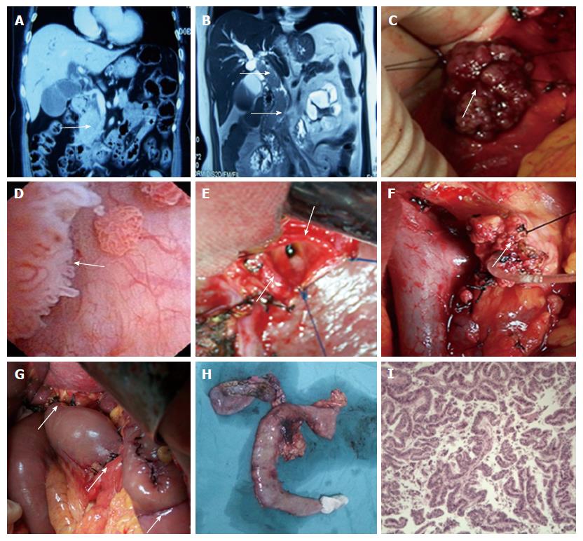
Figure 1 Pancreatoduodenectomy in a 73-year old female patient with bile duct carcinoma.
A: Three-dimensional computed tomography reconstruction found an extrahepatic bile duct obstruction (arrow); B: MRI coronal image confirmed entire extrahepatic bile duct obstruction (arrows); C: Tumor (arrow) seen at the incision of the common bile duct; D: Tumor (arrow) seen in the common bile duct by intraoperative choledochoscopy; E: Upper cut end of the hepatic common duct (arrows); F: The cut end of the pancreas (arrow), and the portal vein; G: Child anastomosis (arrows); H: Resected specimen following pancreatoduodenectomy; I: Papillary adenocarcinoma of extrahepatic bile duct, including the hepatic common duct, common bile duct and Vater ampulla. Hematoxylin and eosin staining. Objective magnification, × 40.
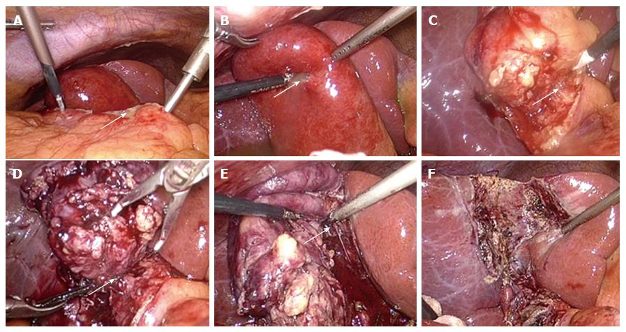
Figure 2 Laparoscopic cholecystectomy in a 78-year-old male patient with acute calculous cholecystitis.
A: Pus (arrow) surrounding the gallbladder; B: Pus (arrow) in the lumen of the gallbladder; C: Congestion and edema of the gallbladder wall (arrow); D: Ordinary silk thread to ligate cystic duct (arrow); E: Inflammatory adhesion (arrow) of gallbladder bed; F: Wound after cholecystectomy.
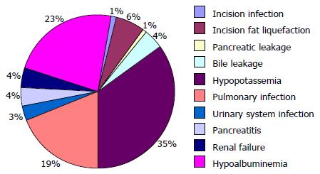
Figure 3 Post-operative complications occurred in 68 elderly patients with biliary diseases.
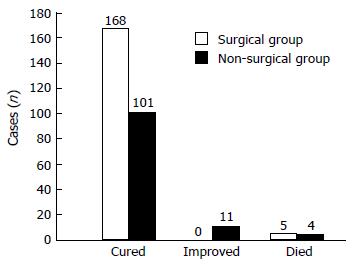
Figure 4 Therapeutic outcomes in the surgical and non-surgical groups.
- Citation: Zhang ZM, Liu Z, Liu LM, Zhang C, Yu HW, Wan BJ, Deng H, Zhu MW, Liu ZX, Wei WP, Song MM, Zhao Y. Therapeutic experience of 289 elderly patients with biliary diseases. World J Gastroenterol 2017; 23(13): 2424-2434
- URL: https://www.wjgnet.com/1007-9327/full/v23/i13/2424.htm
- DOI: https://dx.doi.org/10.3748/wjg.v23.i13.2424


