Copyright
©The Author(s) 2017.
World J Gastroenterol. Mar 21, 2017; 23(11): 2002-2011
Published online Mar 21, 2017. doi: 10.3748/wjg.v23.i11.2002
Published online Mar 21, 2017. doi: 10.3748/wjg.v23.i11.2002
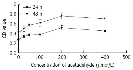
Figure 1 Effects of acetaldehyde for 24 or 48 h on HSC-T6 cell proliferation.
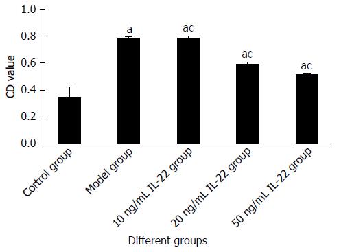
Figure 2 Effects of interleukin-22 on HSC-T6 cells proliferation.
aP < 0.05 vs the control group; cP < 0.05 vs the model group. HSCs: Hepatic stellate cells; IL: Interleukin.
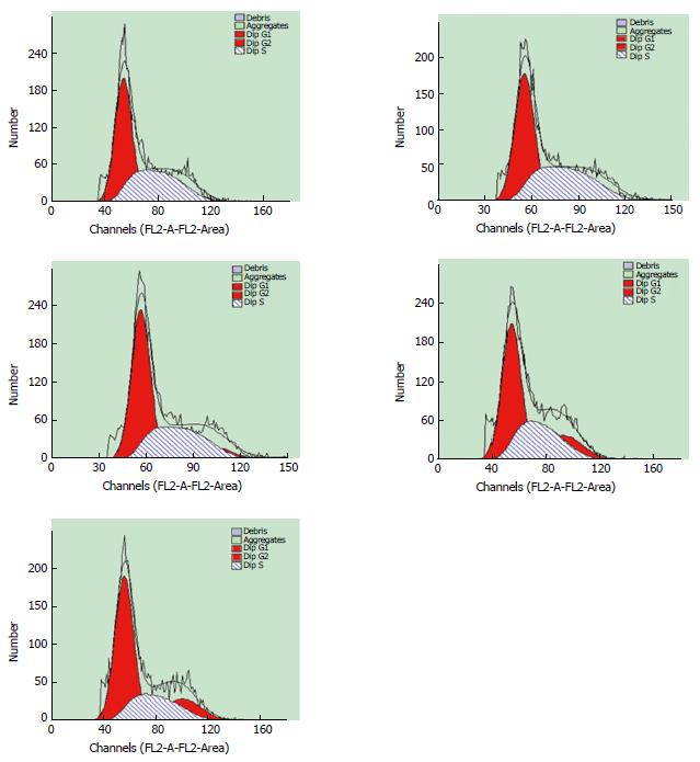
Figure 3 Effects of interleukin-22 on the cell cycle distribution of HSC-T6 cells.
HSCs: Hepatic stellate cells; IL: Interleukin.
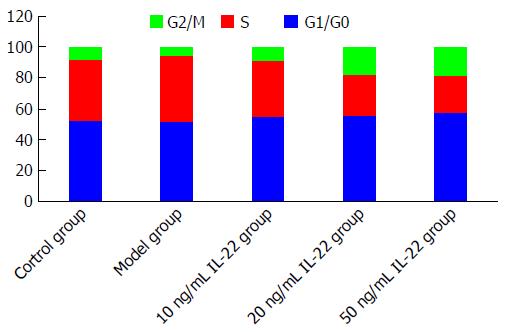
Figure 4 Cell cycle distribution of HSC-T6 cells in different groups.
HSCs: Hepatic stellate cells.
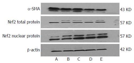
Figure 5 Effects of interleukin-22 on expression of α-smooth muscle antigen and nuclear-factor-related factor 2 protein in acetaldehyde-induced HSC-T6 cells.
A: Control group; B: Model group; C: 10 ng/mL IL-22 group; D: 20 ng/mL IL-22 group; E: 50 ng/mL IL-22 group. IL: Interleukin; SMA: Smooth muscle antigen; Nrf2: Nuclear-factor-related factor 2.
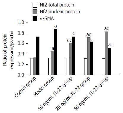
Figure 6 Effects of interleukin-22 on the expression of target protein.
aP < 0.05 vs the control group; cP < 0.05 vs the model group. IL: Interleukin; Nrf2: Nuclear-factor-related factor 2; α-SMA: α-smooth muscle antigen.
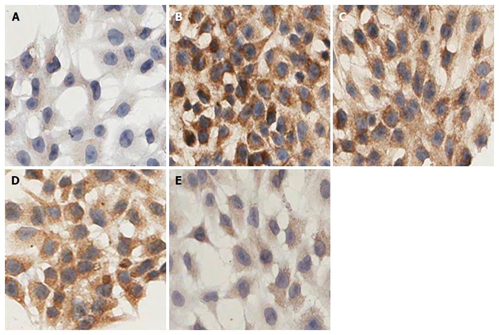
Figure 7 Effects of interleukin-22 on expression of α-smooth muscle antigen (magnification × 400).
A: Control group; B: Model group; C: 10 ng/mL IL-22 group; D: 20 ng/mL IL-22 group; E: 50 ng/mL IL-22 group. All HSC-T6 cells with α-SMA positive expression had brown granules in the cytoplasm, however, the degree of staining varied. In the control group, the cytoplasm was stained light brown, whereas it was obviously deepened in the model group. When treated with different concentrations of IL-22, the staining in the cytoplasm was gradually reduced in a dose-dependent manner. IL: Interleukin; SMA: Smooth muscle antigen.
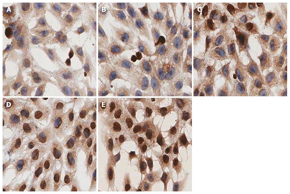
Figure 8 Effects of interleukin-22 on nuclear translocation of nuclear-factor-related factor 2.
A: Control group; B: Model group; C: 10 ng/mL IL-22 group; D: 20 ng/mL IL-22 group; E: 50 ng/mL IL-22 group. Magnification: × 400. IL: Interleukin; SMA: Smooth muscle antigen.
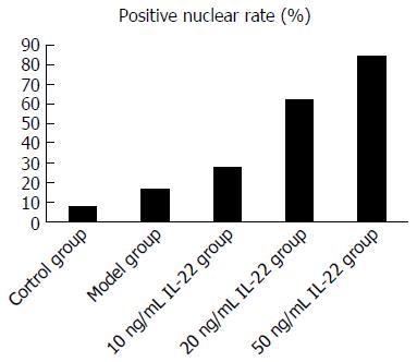
Figure 9 Effects of interleukin-22 on positive rate of nuclear-factor-related factor 2 nuclear staining.
IL: Interleukin.
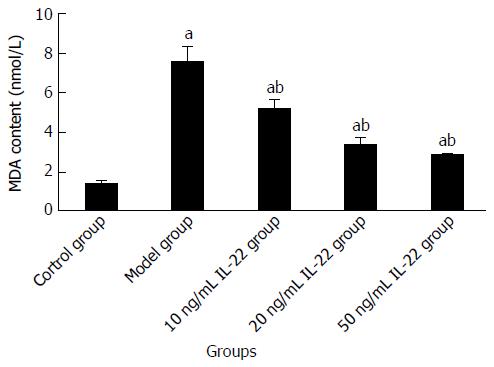
Figure 10 Content of malondialdehyde in different groups.
aP < 0.05 vs the control group; bP < 0.05 vs the model group. MDA: Malondialdehyde; IL: Interleukin.
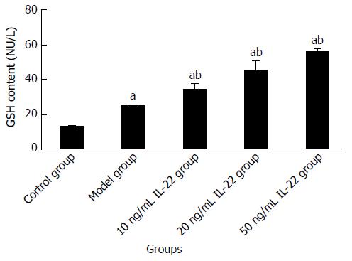
Figure 11 Content of glutathione in different groups.
aP < 0.05 vs the control group; bP < 0.05 vs the model group. GSH: Glutathione; IL: Interleukin.
- Citation: Ni YH, Huo LJ, Li TT. Antioxidant axis Nrf2-keap1-ARE in inhibition of alcoholic liver fibrosis by IL-22. World J Gastroenterol 2017; 23(11): 2002-2011
- URL: https://www.wjgnet.com/1007-9327/full/v23/i11/2002.htm
- DOI: https://dx.doi.org/10.3748/wjg.v23.i11.2002


