Copyright
©The Author(s) 2016.
World J Gastroenterol. Feb 21, 2016; 22(7): 2349-2356
Published online Feb 21, 2016. doi: 10.3748/wjg.v22.i7.2349
Published online Feb 21, 2016. doi: 10.3748/wjg.v22.i7.2349
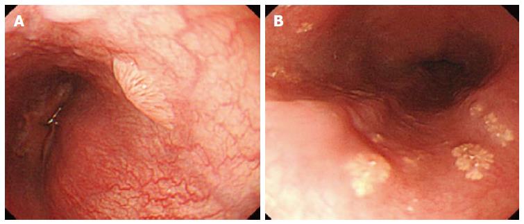
Figure 1 Color of the squamous papilloma of the esophagus looks pale compared to the normal pinkish esophageal mucosa under either near or far focus (A), and the color of the xanthoma looks yellowish under conventional endoscopy (B).
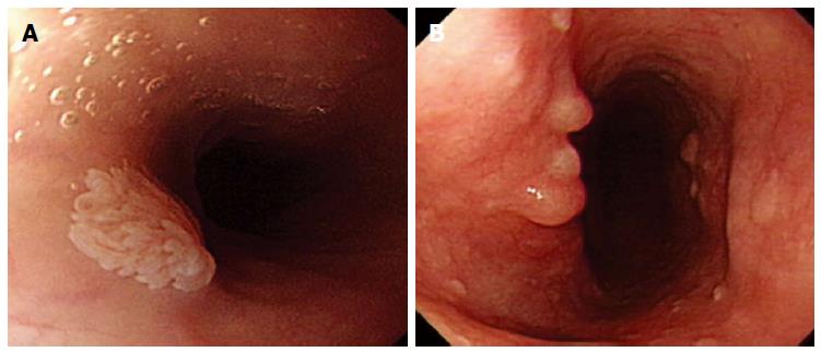
Figure 2 Exophytic growth of squamous papilloma of the esophagus was defined as a lesion that not only bulged from the flat mucosa of the oesophagus but also floated on the plane without complete attachment to the mucosa (A), and SPE may also appear as a sessile polypoid lesion tightly connected to the mucosa (B).
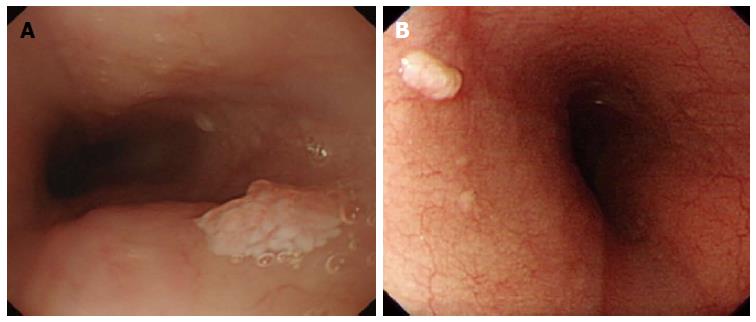
Figure 3 Wart-like shape of squamous papilloma in the esophagus was defined as a lesion with an irregular border but with a margin that could be easily distinguished from the normal esophageal mucosa (A), and the border of squamous hyperplasia was also well-defined and the margin was clear (B).
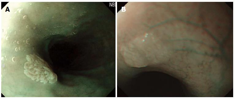
Figure 4 Under UBI mode of endoscopy, vessel crossing was defined as brownish lines passing through the surface of the lesion (A), and by contrast, no brownish lines on glycogenic acanthosis using NBI was observed (B).
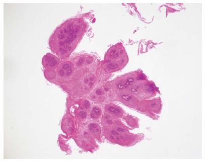
Figure 5 Squamous papilloma of the esophagus is diagnosed pathologically by a hematoxylin and eosin stain.
The lesion is composed of papillary fronds lined by several layers of non-keratinizing squamous epithelium and forming finger-like projections (magnification × 20).
- Citation: Wong MW, Bair MJ, Shih SC, Chu CH, Wang HY, Wang TE, Chang CW, Chen MJ. Using typical endoscopic features to diagnose esophageal squamous papilloma. World J Gastroenterol 2016; 22(7): 2349-2356
- URL: https://www.wjgnet.com/1007-9327/full/v22/i7/2349.htm
- DOI: https://dx.doi.org/10.3748/wjg.v22.i7.2349


