Copyright
©The Author(s) 2016.
World J Gastroenterol. Feb 7, 2016; 22(5): 1844-1853
Published online Feb 7, 2016. doi: 10.3748/wjg.v22.i5.1844
Published online Feb 7, 2016. doi: 10.3748/wjg.v22.i5.1844
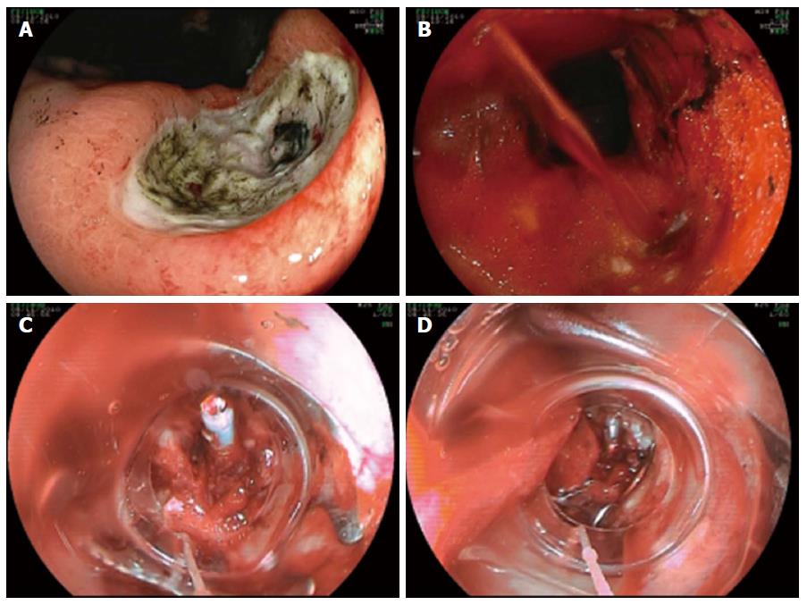
Figure 1 Ulcer (Forrest IIa) at the gastric angulus (A-D).
Acute bleeding after attempt of closure of the visible vessel using a standard hemoclip. Successful hemostasis using a 12/6t over-the-scope-clip.
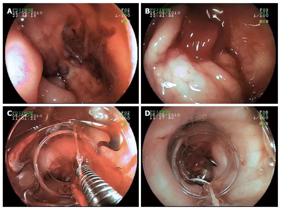
Figure 2 Severe recurrent ulcer bleeding from the gastroduodenal artery at the posterior wall of the duodenal bulb despite primary endoscopic clipping and surgical oversewing (A-D).
Visible surgical threads at the ulcer base (A); Intermittent massive re-bleeding at rinsing and suction (B); Successful application of a 12/6t Over-the-Scope-Clip by pulling the ulcer bed into the transparent distal attachment cap (C, D).
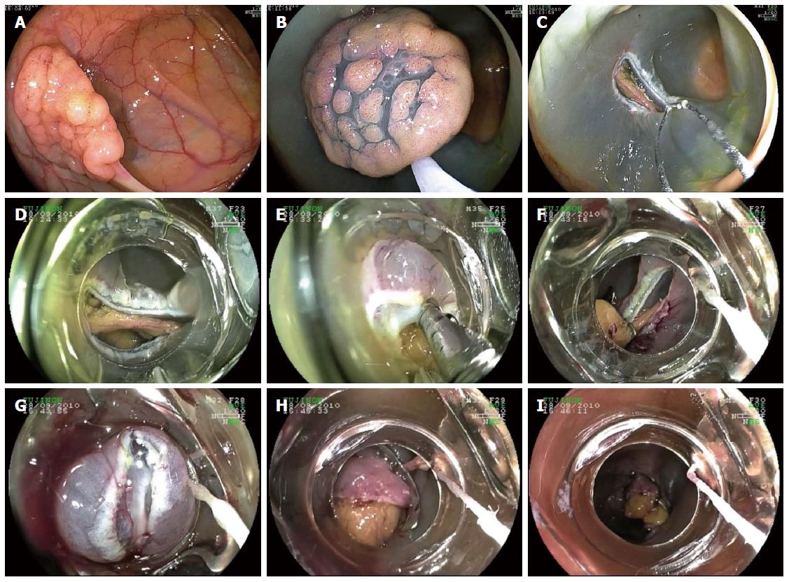
Figure 3 Acute perforation after endoscopic mucosal resection of a 2 cm tubulovillous adenoma at the right colonic flexure (A-I).
Successful closure of the large defect using two large 14/6t over-the-scope-clips applied side-to-side.
- Citation: Wedi E, Gonzalez S, Menke D, Kruse E, Matthes K, Hochberger J. One hundred and one over-the-scope-clip applications for severe gastrointestinal bleeding, leaks and fistulas. World J Gastroenterol 2016; 22(5): 1844-1853
- URL: https://www.wjgnet.com/1007-9327/full/v22/i5/1844.htm
- DOI: https://dx.doi.org/10.3748/wjg.v22.i5.1844


