Copyright
©The Author(s) 2016.
World J Gastroenterol. Feb 7, 2016; 22(5): 1756-1766
Published online Feb 7, 2016. doi: 10.3748/wjg.v22.i5.1756
Published online Feb 7, 2016. doi: 10.3748/wjg.v22.i5.1756
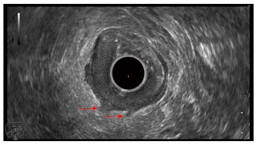
Figure 1 Endoscopic ultrasonography image in a T3 sigmoid cancer showing hypoechoic infiltration beyond the muscularis propria (arrows).
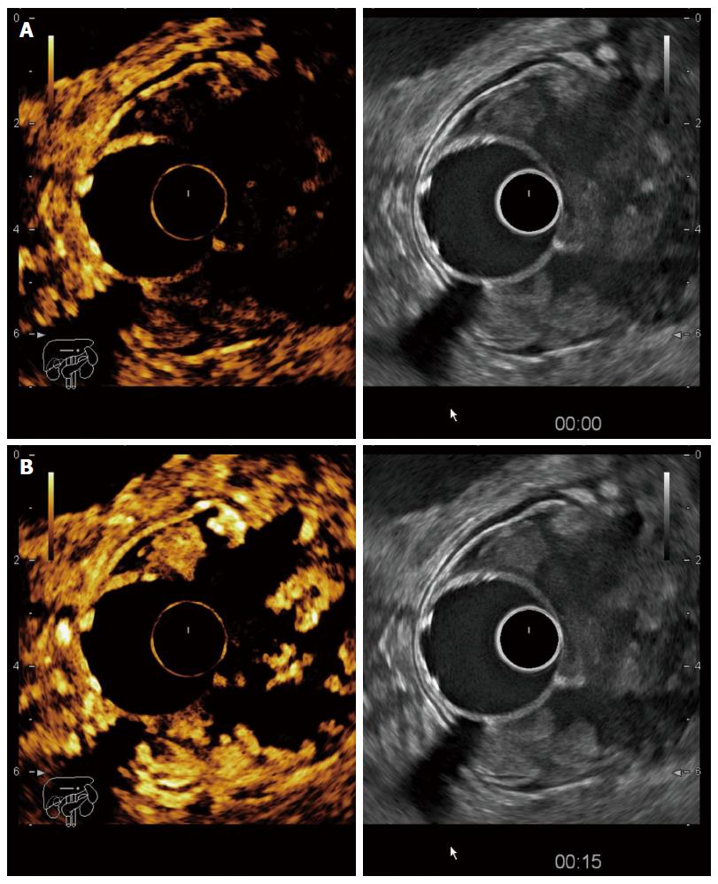
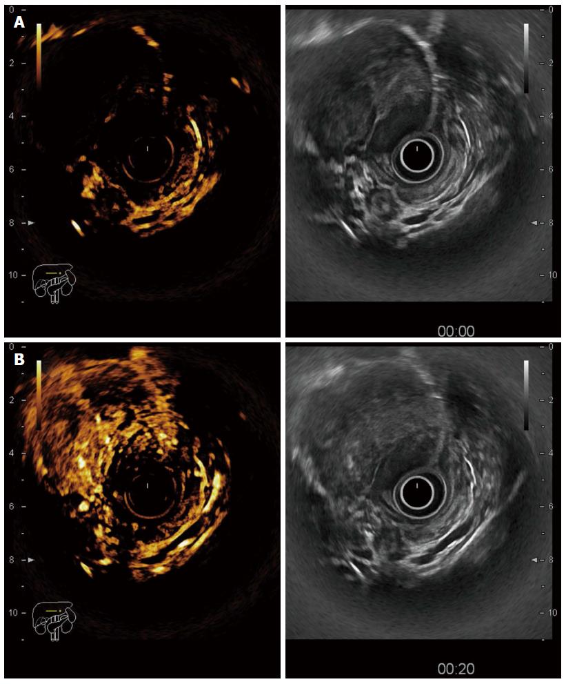
Figure 2 Contrast-enhanced endoscopic ultrasonography in a T3 tumour of the recto-sigmoid junction.
A: Before contrast arrival (left side contrast harmonic imaging mode, right side B mode); B: Maximal enhancement of the tumour 15 s after contrast injection with hyperenhanced areas alternating with avascular (necrotic) areas.
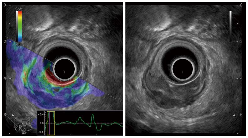
Figure 3 Endoscopic ultrasonography elastography image of a rectal adenocarcinoma with a predominantly blue pattern indicating a low strain mass (left side real-time sono-elastography mode, right side B mode).
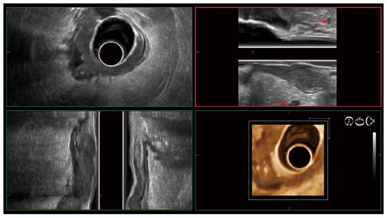
Figure 4 Three-dimensional endoscopic ultrasonography in a T3 rectal cancer with peritumoral lymph nodes (red arrows).
Figure 5 Contrast-enhanced endoscopic ultrasonography in a large rectal gastrointestinal stromal tumour.
A: Before contrast uptake; B: Heterogeneous enhancement after contrast injection.
- Citation: Cârțână ET, Gheonea DI, Săftoiu A. Advances in endoscopic ultrasound imaging of colorectal diseases. World J Gastroenterol 2016; 22(5): 1756-1766
- URL: https://www.wjgnet.com/1007-9327/full/v22/i5/1756.htm
- DOI: https://dx.doi.org/10.3748/wjg.v22.i5.1756


