Copyright
©The Author(s) 2016.
World J Gastroenterol. Dec 21, 2016; 22(47): 10471-10476
Published online Dec 21, 2016. doi: 10.3748/wjg.v22.i47.10471
Published online Dec 21, 2016. doi: 10.3748/wjg.v22.i47.10471
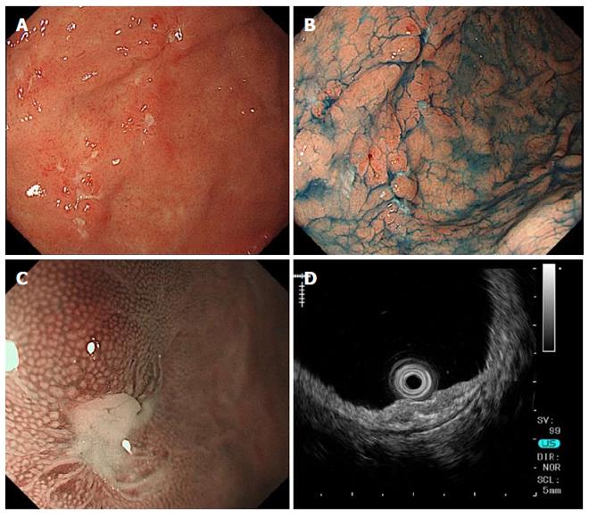
Figure 1 Gastroendoscopy findings before corticosteroid treatment.
A, B: Multiple map-like ulcerative lesions and ulcer scars are seen on the greater curvature of the body to the fornix and atrophic gastritis with Helicobacter pylori infection; C: Magnifying narrow-band imaging shows the absence of epithelial change at the edge of the ulcer; D: Endoscopic ultrasonography reveals thickness and irregularity in the second layer of the gastric wall.
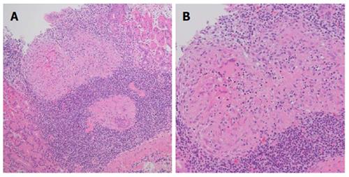
Figure 2 Histopathologic findings of the gastric mucosal biopsy specimen before corticosteroid treatment.
The mucosal layer of the gastric wall shows multiple noncaseating granulomas and infiltration of inflammatory cells, especially lymphocytes. A: HE, magnification × 10; B: HE, magnification × 20. HE: Hematoxylin and eosin.
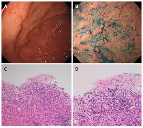
Figure 3 Gastroendoscopy and histopathologic findings after concomitant treatment with corticosteroid and azathioprine.
A, B: Most of the sarcoidosis-related gastric ulcers in the fornix are healed; C, D: Gastric mucosal granuloma and lymphocytic infiltration is decreased; C: HE, magnification × 10; D: HE, magnification × 20. HE: Hematoxylin and eosin.
- Citation: Murata M, Sugimoto M, Yokota Y, Ban H, Inatomi O, Bamba S, Kushima R, Andoh A. Efficacy of additional treatment with azathioprine in a patient with prednisolone-dependent gastric sarcoidosis. World J Gastroenterol 2016; 22(47): 10471-10476
- URL: https://www.wjgnet.com/1007-9327/full/v22/i47/10471.htm
- DOI: https://dx.doi.org/10.3748/wjg.v22.i47.10471


