Copyright
©The Author(s) 2016.
World J Gastroenterol. Dec 21, 2016; 22(47): 10380-10387
Published online Dec 21, 2016. doi: 10.3748/wjg.v22.i47.10380
Published online Dec 21, 2016. doi: 10.3748/wjg.v22.i47.10380
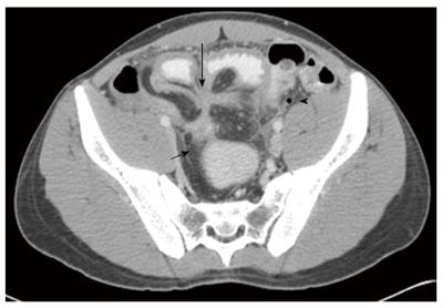
Figure 1 Axial, post contrast computed tomography (image of a 21 yr male with first time presentation of Crohn's disease.
There is approximately 20 cm distal ileal bowel segment involved in the disease with wall thickening and increased enhancement (level 2). There are 2 fistula formation; one entero-enteric between the distal small bowel and terminal ileum (long arrow) and second, entero-colonic fistula involving the sigmoid bowel (short arrow). The oral contrast material has not reached the colon at the time of the scan, however it is seen in the sigmoid due to passage through the entero-colonic fistula. A small amount of free fluid is seen on the left with a free air bubble from suspected perforation (arrowhead). There is also pre stenotic dilatation proximal to the fistulas, not seen in the image.
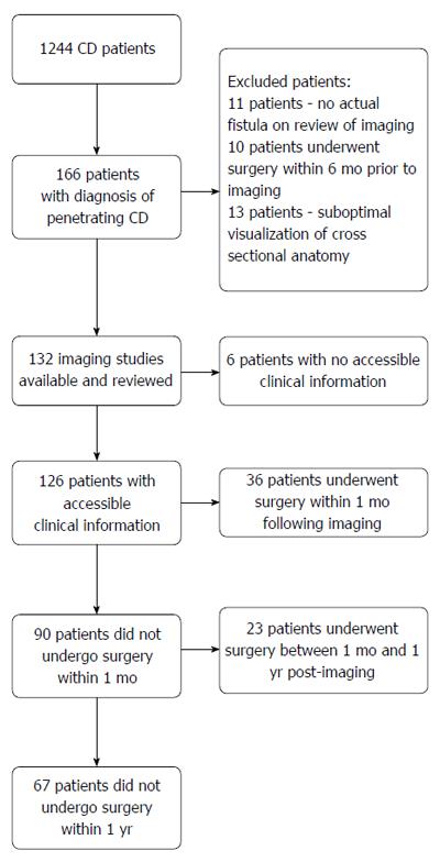
Figure 2 Flow diagram of enrolment and patient outcomes.
CD: Crohn’s disease.
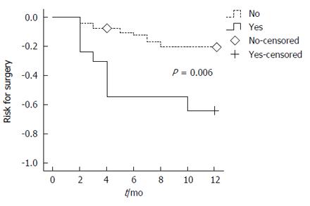
Figure 3 Survival rate free of resection-surgery.
In this Kaplan-Meier analysis, the rate free of resection-surgery is shown. High risk (solid line) - more than one fistula or stricture or entero-vesical fistula. Low risk (dashed line) - none of the above.
- Citation: Yaari S, Benson A, Aviran E, Lev Cohain N, Oren R, Sosna J, Israeli E. Factors associated with surgery in patients with intra-abdominal fistulizing Crohn's disease. World J Gastroenterol 2016; 22(47): 10380-10387
- URL: https://www.wjgnet.com/1007-9327/full/v22/i47/10380.htm
- DOI: https://dx.doi.org/10.3748/wjg.v22.i47.10380


