Copyright
©The Author(s) 2016.
World J Gastroenterol. Dec 21, 2016; 22(47): 10316-10324
Published online Dec 21, 2016. doi: 10.3748/wjg.v22.i47.10316
Published online Dec 21, 2016. doi: 10.3748/wjg.v22.i47.10316
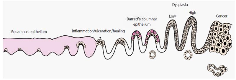
Figure 1 Progression from normal squamous epithelium to oesophageal cancer in Barrett’s oesophagus[76].
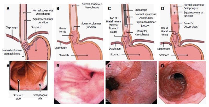
Figure 2 Despite these recognised anatomical landmarks documentation is challenging and there is the potential for inter and intra-observer variation.
A: Normal oesophagus and stomach; B: Hiatus Hernia; C: Short segment Barrett’s; D: Long segment Barrett’s[77].
- Citation: Evans RPT, Mourad MM, Fisher SG, Bramhall SR. Evolving management of metaplasia and dysplasia in Barrett's epithelium. World J Gastroenterol 2016; 22(47): 10316-10324
- URL: https://www.wjgnet.com/1007-9327/full/v22/i47/10316.htm
- DOI: https://dx.doi.org/10.3748/wjg.v22.i47.10316


