Copyright
©The Author(s) 2016.
World J Gastroenterol. Dec 7, 2016; 22(45): 9944-9953
Published online Dec 7, 2016. doi: 10.3748/wjg.v22.i45.9944
Published online Dec 7, 2016. doi: 10.3748/wjg.v22.i45.9944
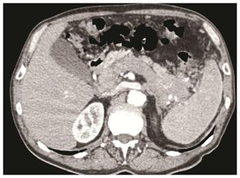
Figure 1 Pancreatic poorly differentiated neuroendocrine carcinomas of the pancreatic body-tail associated with neoplastic thrombosis of the splenic vein/portal vein, and with a lymphadenopathy along the stomach.
The patients underwent left pancreatectomy with splenectomy, portal vein resection with portal vein thrombectomy and partial gastric resection.
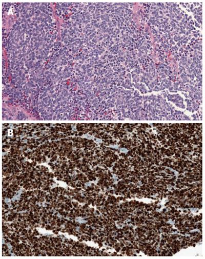
Figure 2 A small cell poorly differentiated neuroendocrine carcinoma of the pancreas (A) (haematoxylin-eosin stain) and a small cell poorly differentiated neuroendocrine carcinoma of the pancreas with high ki67 proliferative index (B, Ki67: 90%).
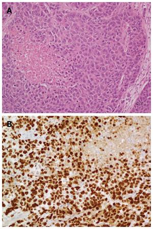
Figure 3 Shows a large cell poorly differentiated neuroendocrine carcinoma of the pancreas (A, haematoxylin-eosin stain) and a large cell poorly differentiated neuroendocrine carcinoma of the pancreas with its Ki67 proliferative index (B, Ki67: 80%).
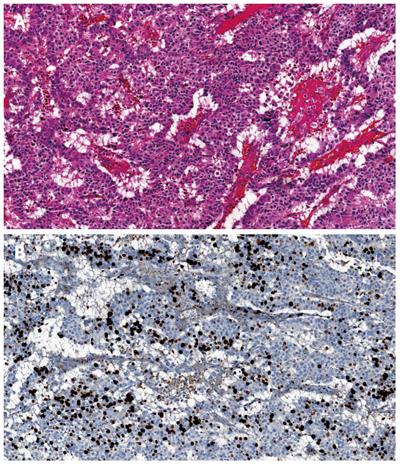
Figure 4 Morphological well differentiated neuroendocrine carcinoma of the pancreas (A, haematoxylin-eosin stain) and morphological well differentiated neuroendocrine carcinoma of the pancreas with Ki67 proliferative index of 30% (B).
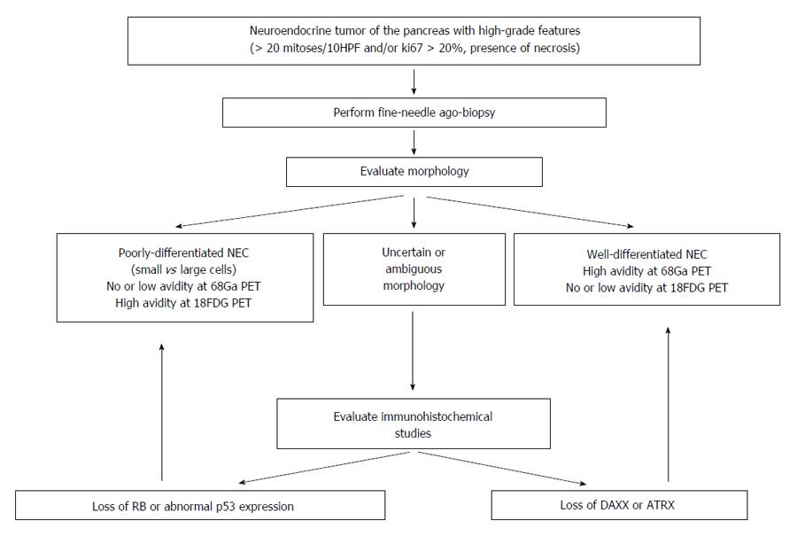
Figure 5 Diagnostic flowchart algorithm in patients with pancreatic neuroendocrine carcinomas.
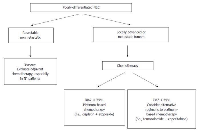
Figure 6 Different therapeutic options in patients with morphological poorly differentiated neuroendocrine carcinomas of the pancreas.
18FDG-PET: 18F-fluorodeoxyglucose positron emission tomography.
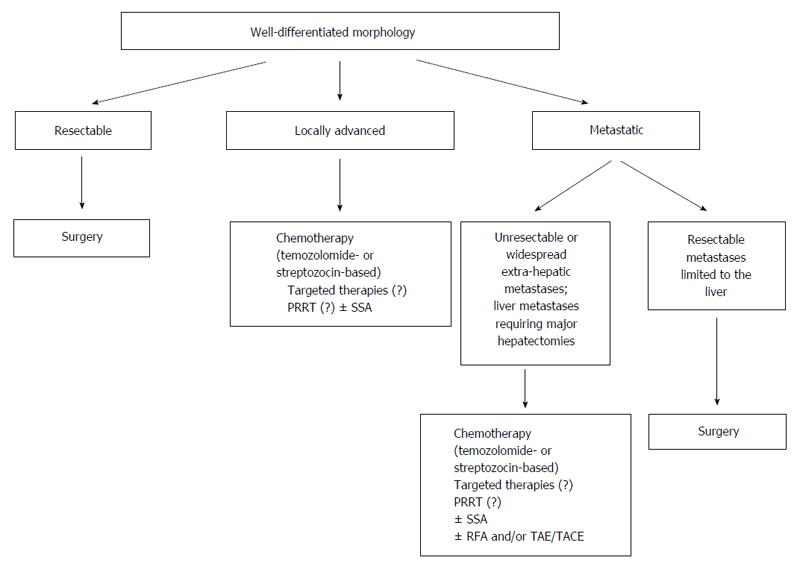
Figure 7 Management flowchart algorithm in patients with morphological well differentiated neuroendocrine carcinomas of the pancreas.
PRRT: Peptide receptor radionuclide therapy; TAE: Transarterial embolization; TACE: Transarterial chemoembolization; SSA: Somatostatin analogues.
- Citation: Crippa S, Partelli S, Belfiori G, Palucci M, Muffatti F, Adamenko O, Cardinali L, Doglioni C, Zamboni G, Falconi M. Management of neuroendocrine carcinomas of the pancreas (WHO G3): A tailored approach between proliferation and morphology. World J Gastroenterol 2016; 22(45): 9944-9953
- URL: https://www.wjgnet.com/1007-9327/full/v22/i45/9944.htm
- DOI: https://dx.doi.org/10.3748/wjg.v22.i45.9944


