Copyright
©The Author(s) 2016.
World J Gastroenterol. Dec 7, 2016; 22(45): 9909-9920
Published online Dec 7, 2016. doi: 10.3748/wjg.v22.i45.9909
Published online Dec 7, 2016. doi: 10.3748/wjg.v22.i45.9909
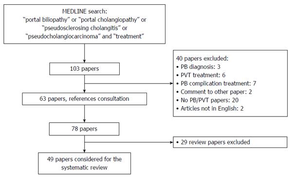
Figure 1 Flow-chart of literature search.
PB: Portal biliopathy; PVT: Portal vein thrombosis.
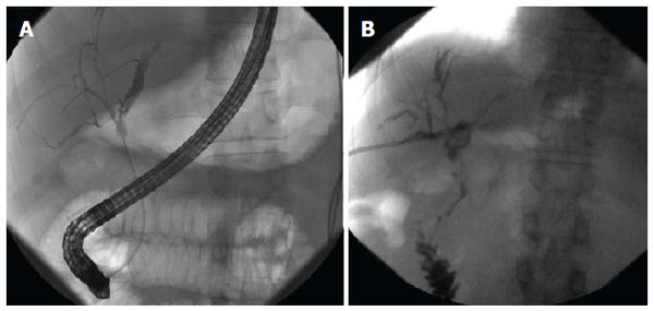
Figure 2 Cholangiographic findings in a patient with symptomatic (jaundice and cholangitis) portal biliopathy secondary to chronic extrahepatic portal vein obstruction and portal cavernoma.
Ischemic stenosis with dilatation of the left intrahepatic biliary tree is shown by ERCP (A): patient underwent unsuccessful ERCP with stent insertion and then PTBD placement; PTBD was changed for 3 times in one year because of cholangitis and liver abscess. After clinical and biochemical improvement of BA, patient was treated with surgical splenorenal shunt and after 5 mo PTBD was definitively removed. Last cholangiography obtained before PTBD removal shows significant improvement in biliary dilation (B). Patient is actually asymptomatic for BA and PB management. BA: Bilioenteric anastomosis; ERCP: Endoscopic retrograde cholangiopancreatography; PB: Portal biliopathy; PTBD: Percutaneous transhepatic biliary drainage.
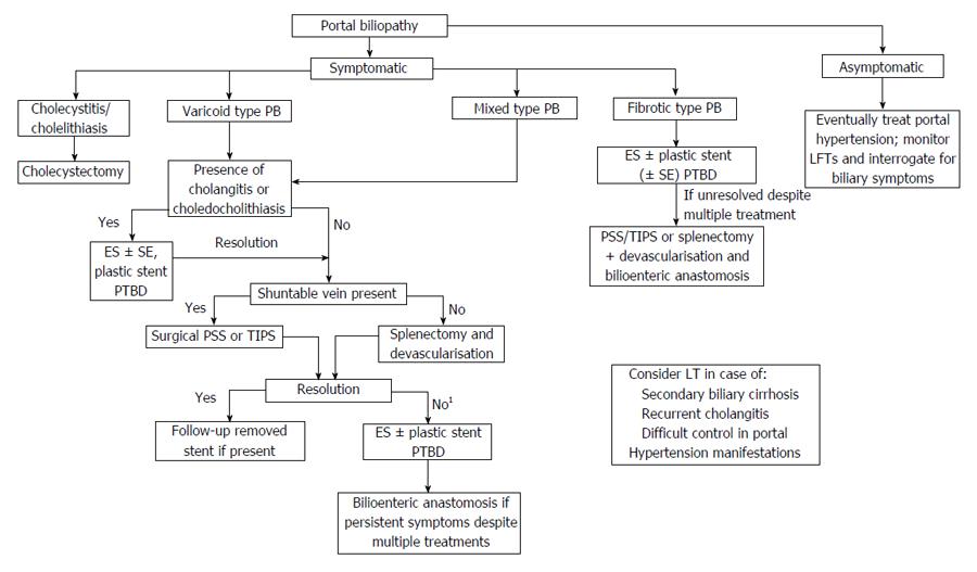
Figure 3 Proposed algorithm for portal biliopathy management.
1Unsuccessful vascular surgery is frequent in mixed type because of co-presence of ischemic and compressive damage. ES: Endoscopic sphincterotomy; SE: Stone extraction; LFTs: Liver function tests; LT: Liver transplantation; PTBD: Percutaneous transhepatic biliary drainage; PB: Portal biliopathy; PSS: Porto-systemic shunt; TIPS: Transjugular intrahepatic porto-systemic shunt.
- Citation: Franceschet I, Zanetto A, Ferrarese A, Burra P, Senzolo M. Therapeutic approaches for portal biliopathy: A systematic review. World J Gastroenterol 2016; 22(45): 9909-9920
- URL: https://www.wjgnet.com/1007-9327/full/v22/i45/9909.htm
- DOI: https://dx.doi.org/10.3748/wjg.v22.i45.9909


