Copyright
©The Author(s) 2016.
World J Gastroenterol. Nov 28, 2016; 22(44): 9661-9673
Published online Nov 28, 2016. doi: 10.3748/wjg.v22.i44.9661
Published online Nov 28, 2016. doi: 10.3748/wjg.v22.i44.9661
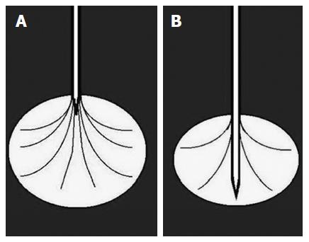
Figure 1 Needle with expandable electrodes.
Electrodes can be opened within the lesion from the top (A) or from the back (B) of the needle.
Figure 2 Needle with single electrode.
Single electrode of the needle within the lesion.
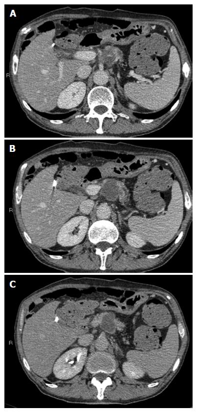
Figure 3 Radiofrequency ablation of pancreatic cancer.
Computed tomography (CT) scan in the portal phase (A, B) shows the markedly hypodense necrotic avascular area modelled within the tumor. CT scan in the late phase (C) shows the ablated area as being better delineated from the enhanced adjacent tissue.
- Citation: D’Onofrio M, Ciaravino V, De Robertis R, Barbi E, Salvia R, Girelli R, Paiella S, Gasparini C, Cardobi N, Bassi C. Percutaneous ablation of pancreatic cancer. World J Gastroenterol 2016; 22(44): 9661-9673
- URL: https://www.wjgnet.com/1007-9327/full/v22/i44/9661.htm
- DOI: https://dx.doi.org/10.3748/wjg.v22.i44.9661


