Copyright
©The Author(s) 2016.
World J Gastroenterol. Nov 21, 2016; 22(43): 9562-9570
Published online Nov 21, 2016. doi: 10.3748/wjg.v22.i43.9562
Published online Nov 21, 2016. doi: 10.3748/wjg.v22.i43.9562
Figure 1 Small paraductal cysts in the pancreatic hystmus on baseline magnetic resonance cholangiopancreatography in a 65 years old female patient (A), showing increase in size at 24 mo (B) and then decrease in size at 36 mo (C).
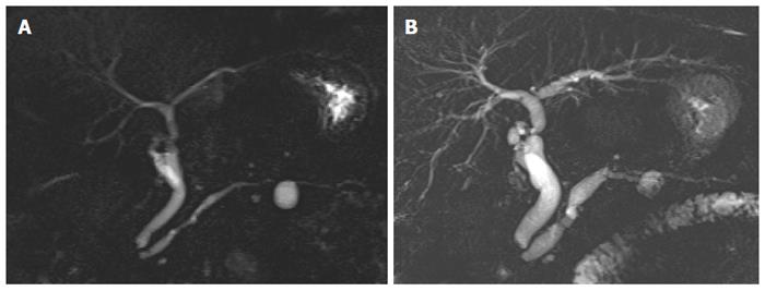
Figure 2 Occurrence of alert findings in patient number 5 described in Table 3.
Compared to the baseline examination (A), follow-up MRCP (B) showed main pancreatic duct dilation and small parietal filling defects in the body cyst. MRCP: Magnetic resonance cholangiopancreatography.
- Citation: Girometti R, Pravisani R, Intini SG, Isola M, Cereser L, Risaliti A, Zuiani C. Evolution of incidental branch-duct intraductal papillary mucinous neoplasms of the pancreas: A study with magnetic resonance imaging cholangiopancreatography. World J Gastroenterol 2016; 22(43): 9562-9570
- URL: https://www.wjgnet.com/1007-9327/full/v22/i43/9562.htm
- DOI: https://dx.doi.org/10.3748/wjg.v22.i43.9562


