Copyright
©The Author(s) 2016.
World J Gastroenterol. Nov 7, 2016; 22(41): 9235-9241
Published online Nov 7, 2016. doi: 10.3748/wjg.v22.i41.9235
Published online Nov 7, 2016. doi: 10.3748/wjg.v22.i41.9235
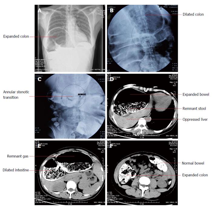
Figure 1 Imaging examinations.
The radiography (A) showed right pleural effusion and thickening, and potential interposition of the dilated colon. The colon double contract pneumobarium radiography (B and C) and the abdominopelvic computed tomography (D-F) revealed significant expansion of the cecum, the ascending colon, the hepatic flexure of colon, and the remnant transverse colon, with the most dilated area 13.6 cm wide. Abundant residual stool, gas, and liquid existed. An annular stenotic transitional segment (only 3.8 cm wide) could be perceived. The other intestines were normal. The neighboring organs and tissues underwent marked displacement and deformation under pressure.
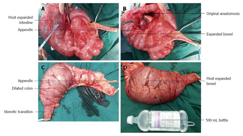
Figure 2 Surgical pictures.
In the second surgery, right colectomy was conducted to remove the aganglionic and the dilated fragments (A and B), and the resected intestine included the non-dilated bowel 6 cm beyond the anastomosis of the initial surgery (C), ensuring the completeness and definitiveness of the removal. The affected colon was obviously outstretched (D).
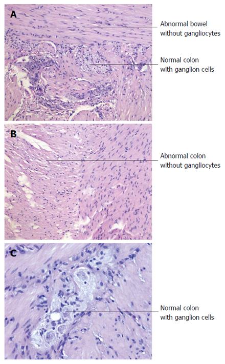
Figure 3 Postsurgical pathological examination.
A: Shows the junction of the normal and the abnormal bowel contained in the resected specimen (magnification × 100); B: Indicates the resected abnormal bowel segment without ganglion cells (magnification × 100). The cutting edge contained abundant normal ganglion cells (C, magnification × 200), ensuring the total and complete removal of the abnormal and aganglioniccolon segment.
- Citation: Wei ZJ, Huang L, Xu AM. Reoperation in an adult female with "right-sided" Hirschsprung's disease complicated by refractory hypertension and cough. World J Gastroenterol 2016; 22(41): 9235-9241
- URL: https://www.wjgnet.com/1007-9327/full/v22/i41/9235.htm
- DOI: https://dx.doi.org/10.3748/wjg.v22.i41.9235


