Copyright
©The Author(s) 2016.
World J Gastroenterol. Oct 28, 2016; 22(40): 8940-8948
Published online Oct 28, 2016. doi: 10.3748/wjg.v22.i40.8940
Published online Oct 28, 2016. doi: 10.3748/wjg.v22.i40.8940
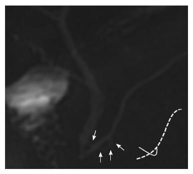
Figure 1 Ansa pancreatica depicted on magnetic resonance images and its schematic image.
Magnetic resonance cholangiopancreatography reveals the absence of a normal type of accessory pancreatic duct and the presence of an additional curved duct in the head of pancreas (white arrows). The additional duct is seen crossing the main pancreatic duct on this projection image. Schematic image of the pancreatic duct is described on the right side. The broken line indicates the ventral duct and the solid black line represents ansa pancreatica.
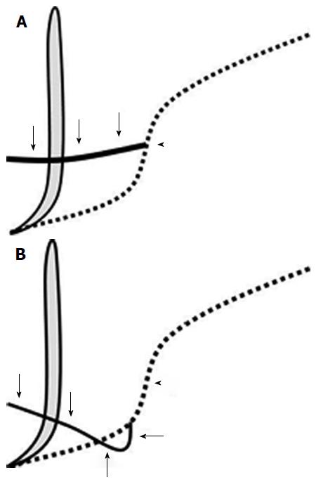
Figure 2 Schematic images of a normal pancreatic duct (A) and ansa pancreatica (B).
The vertical thick gray line indicates the common bile duct and the broken lines indicate the ventral duct. The solid black line represents the normal accessory duct in (A) and ansa pancreatica in (B). In the normal type (A), the accessory duct (arrows) arises near the flexion point (arrowhead) of the main pancreatic duct and runs towards the minor papilla horizontally and to the right. In ansa pancreatica (B), the additional duct (arrows) arises from the caudal side of the flexion point (arrowhead) of the main pancreatic duct. It runs caudally at first, then rightward and ventrally, crossing the ventral duct, and finally terminates near the minor papilla.
- Citation: Hayashi TY, Gonoi W, Yoshikawa T, Hayashi N, Ohtomo K. Ansa pancreatica as a predisposing factor for recurrent acute pancreatitis. World J Gastroenterol 2016; 22(40): 8940-8948
- URL: https://www.wjgnet.com/1007-9327/full/v22/i40/8940.htm
- DOI: https://dx.doi.org/10.3748/wjg.v22.i40.8940


