Copyright
©The Author(s) 2016.
World J Gastroenterol. Oct 21, 2016; 22(39): 8849-8852
Published online Oct 21, 2016. doi: 10.3748/wjg.v22.i39.8849
Published online Oct 21, 2016. doi: 10.3748/wjg.v22.i39.8849
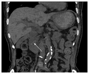
Figure 1 Abdominal computed tomography scan on admission.
There are multiple small computed tomography-high density stones on lower common bile duct (white arrow).
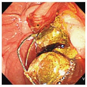
Figure 2 Emergent endoscopic retrograde cholangiopancreatography.
With endoscopic sphincterotome and mechanical lithotripsy, all stones could be removed successfully without any complications of either hemorrhage or gastrointestinal perforation.
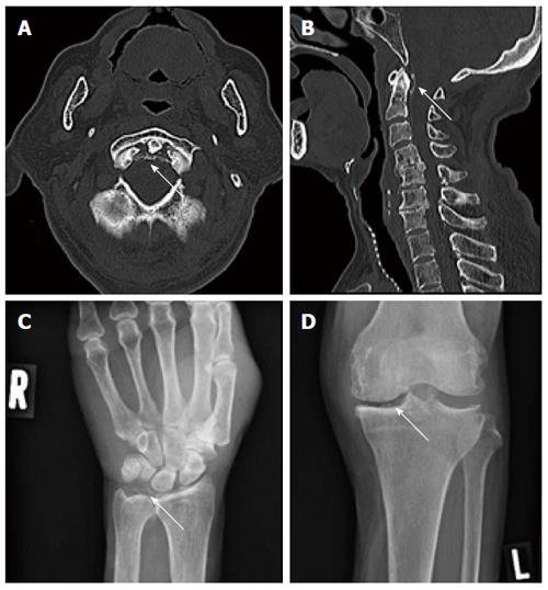
Figure 3 Cervical computed tomography and radiograph of the right wrist and left knee on day 4 after endoscopic retrograde cholangiopancreatography.
A: A sharply defined calcification of periodontoid articular ligaments is noted (white arrow); B: The same as (A) on sagittal view; C: Crystal deposit in the right wrist joint space (white arrow); D: Crystal deposit in left knee joint space (white arrow).
- Citation: Nakano H, Nakahara K, Michikawa Y, Suetani K, Morita R, Matsumoto N, Itoh F. Crowned dens syndrome developed after an endoscopic retrograde cholangiopancreatography procedure. World J Gastroenterol 2016; 22(39): 8849-8852
- URL: https://www.wjgnet.com/1007-9327/full/v22/i39/8849.htm
- DOI: https://dx.doi.org/10.3748/wjg.v22.i39.8849


