Copyright
©The Author(s) 2016.
World J Gastroenterol. Oct 14, 2016; 22(38): 8576-8583
Published online Oct 14, 2016. doi: 10.3748/wjg.v22.i38.8576
Published online Oct 14, 2016. doi: 10.3748/wjg.v22.i38.8576
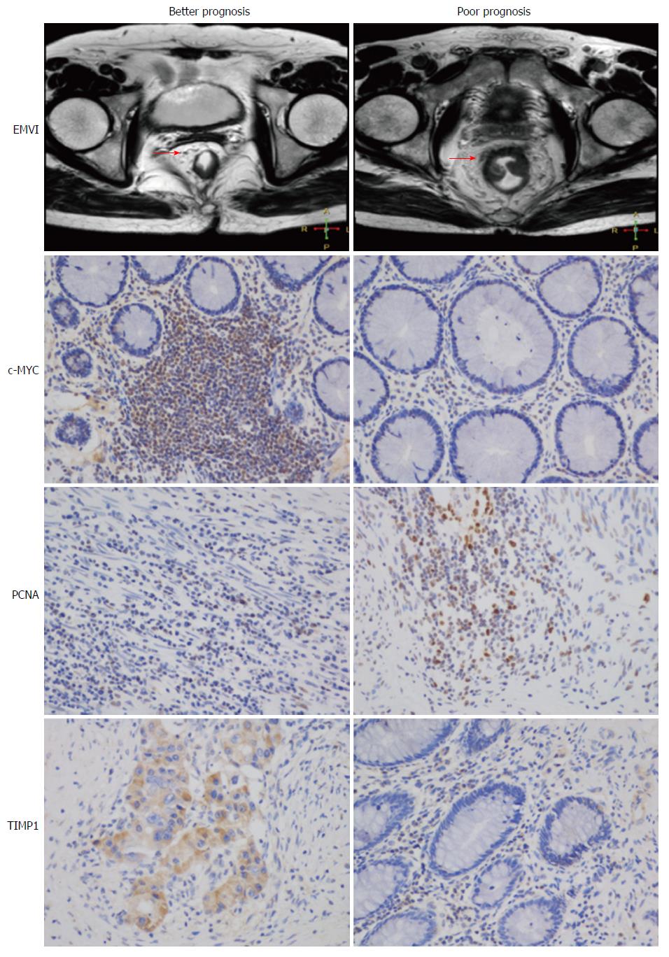
Figure 1 Representative examples of extramural vascular invasion and immunohistochemistry images in distinct prognostic groups.
Overexpression of c-MYC and TIMP1 is a marker of better prognosis, whereas PCNA is a marker of poor prognosis, which was consistent with known biological roles.
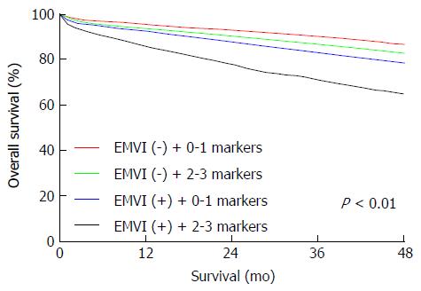
Figure 2 Immunohistochemistry panel combined with magnetic resonance imaging-detected extramural vascular invasion enhanced the accuracy of prognoses for patients with rectal carcinoma.
Combination of EMVI and the IHC panel separated patients into distinct prognostic groups (P < 0.01). EMVI: Extramural vascular invasion; IHC: Immunohistochemistry.
- Citation: Li XF, Jiang Z, Gao Y, Li CX, Shen BZ. Combination of three-gene immunohistochemical panel and magnetic resonance imaging-detected extramural vascular invasion to assess prognosis in non-advanced rectal cancer patients. World J Gastroenterol 2016; 22(38): 8576-8583
- URL: https://www.wjgnet.com/1007-9327/full/v22/i38/8576.htm
- DOI: https://dx.doi.org/10.3748/wjg.v22.i38.8576


