Copyright
©The Author(s) 2016.
World J Gastroenterol. Oct 7, 2016; 22(37): 8375-8381
Published online Oct 7, 2016. doi: 10.3748/wjg.v22.i37.8375
Published online Oct 7, 2016. doi: 10.3748/wjg.v22.i37.8375
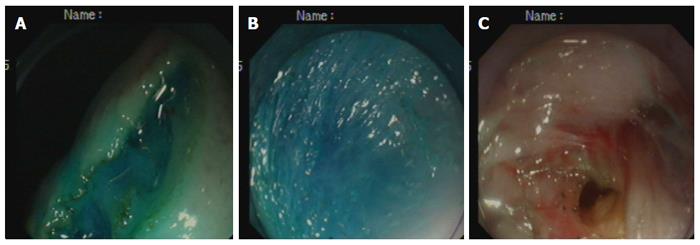
Figure 1 Step-by-step procedure of pelvis-directed submucosal tunneling endoscopic gastrostomy.
A: Gastric mucosal incision; B: Submucosal tunneling; C: Seromuscular incision.
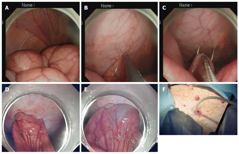
Figure 2 Step-by-step procedure of endoscopic tube ileostomy.
A: Endoscopic identification of the distal ileum in the left lower quadrant; B: Endoscopic view of a loop of the distal ileum on its anti-mesenteric side grasped and held to the anterior abdominal wall; C: Percutaneous terminal ileum puncture; D: Endoscopic view of the distal ileum sutured to the abdominal wall with two stitches; E: Endoscopic view of the inflated balloon in the ileal lumen; F: Accomplished tube ileostomy.
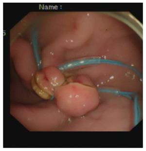
Figure 3 An endoscopic view of gastrostomy closure with endoloop ligation.
Figure 4 Gross and histopathological evaluation of gastrostomy closure 1 wk after natural orifice transgastric endoscopic surgery.
A: Gross examination showing complete healing of gastrostomy; B: Histopathological examination showing complete healing of gastrostomy.
Figure 5 Gross and histopathological evaluation of stoma tract formation of the ileostomy 1 wk after natural orifice transgastric endoscopic surgery.
A: Gross examination showing adequate stoma tract formation of ileostomy; B: Histopathological examination showing adequate stoma tract formation of ileostomy.
- Citation: Shi H, Chen SY, Wang YG, Jiang SJ, Cai HL, Lin K, Xie ZF, Dong FF. Percutaneous transgastric endoscopic tube ileostomy in a porcine survival model. World J Gastroenterol 2016; 22(37): 8375-8381
- URL: https://www.wjgnet.com/1007-9327/full/v22/i37/8375.htm
- DOI: https://dx.doi.org/10.3748/wjg.v22.i37.8375


