Copyright
©The Author(s) 2016.
World J Gastroenterol. Sep 28, 2016; 22(36): 8234-8241
Published online Sep 28, 2016. doi: 10.3748/wjg.v22.i36.8234
Published online Sep 28, 2016. doi: 10.3748/wjg.v22.i36.8234
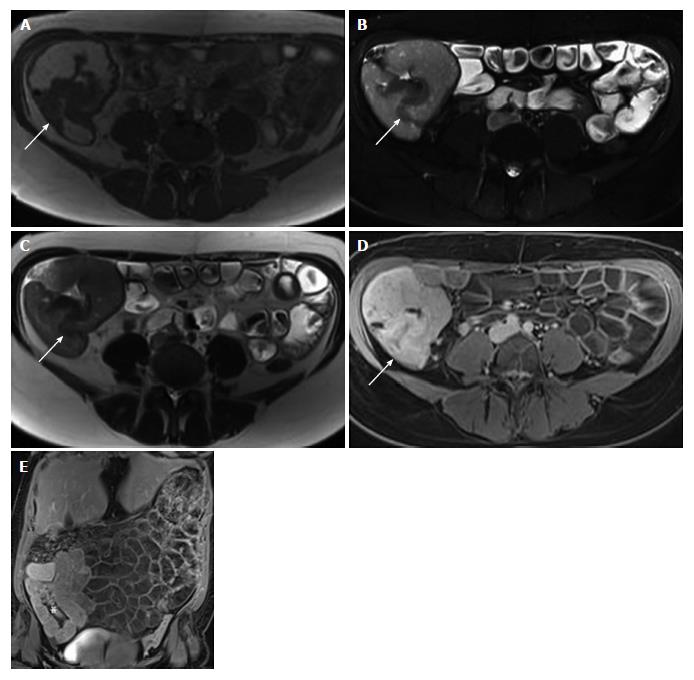
Figure 1 Magnetic resonance imaging showed mural thickening of the ileocecum and the appendix, which was interpreted as being inflammatory in nature.
A-E: Magnetic resonance images (A: T2-weighted magnetization transfer contrast transversal; B: T2-weighted fat saturated transversal; C: T2-weighted half-Fourier acquired single-shot turbo spin-echo transversal; D: T1-weighted VIBE-Dixon postcontrast transversal, E: T1-weighted VIBE-Dixon postcontrast coronal) showing mural thickening (arrows in A-C) of the ileocecum and the appendix with contrast enhancement (arrow in D, asterix in E).
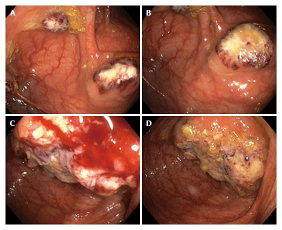
Figure 2 Colonoscopy.
A, B: Colonoscopy showing multiple pseudotumors (amebomas) in the ascending colon, mimicking carcinoma; C, D: Endoscopic view of a large ulcerated tumor mass at the ileocecal valve.
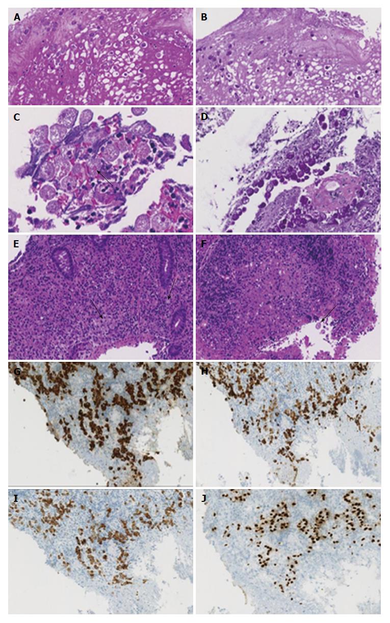
Figure 3 Microscopic examination.
A, B: Microscopy (A: HE stain, × 20, B: PAS, × 20) of biopsy specimens from the ascending colon showing aggregations of round PAS-positive Entamoeba histolytica trophozoites within cell debris; C, D: High power view (C: HE stain, × 40, D: PAS, × 20) demonstrating PAS-positive trophozoites containing ingested erythrocytes (arrow in C); E, F: Microscopy (HE stain, × 20) of biopsy specimens from the ileocecal valve showing infiltrating signet-ring cells (arrows in E) in combination with amebic protozoa (arrow in F); G-J: The tumor cells stain positive with pancytokeratin (G: × 20), CK 7 (H: × 20), CK 20 (I: × 20) and CDX-2 (J: × 20).
Figure 4 Explorative laparoscopy.
A, B: Laparoscopic view of a tumor mass (arrows) at the ileocecum and the appendix.
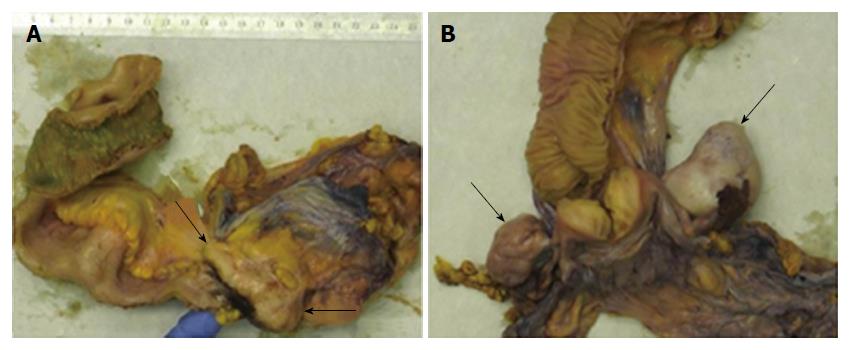
Figure 5 Gross pathology of the surgical specimens.
A: Gross image showing a tumor mass (arrows) that involves the appendix, the ileocecal valve and the cecum. Surgical resection was performed after eradication of amebiasis and chemotherapy. B: Gross image of the Douglas-resection showing bilateral ovary metastases (arrows).
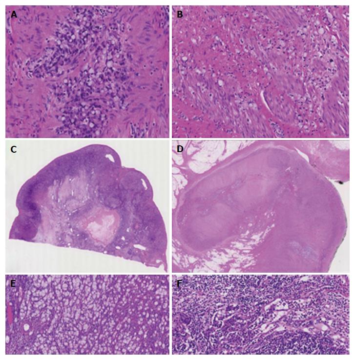
Figure 6 Biopsy-based diagnosis of signet-ring cell adenocarcinoma was confirmed by histology of the resected specimens.
A, B: Microscopy (A: HE stain, × 40, B: HE stain, × 20) of surgical specimens demonstrating infiltration of the colonic wall by signet-ring cells; C, D: The tumor involves the cecum (C: HE stain, × 4) and the appendix (D: HE stain, × 4); E, F: Microscopy showing metastases to the ovary (D: HE stain, × 10) and lymph nodes (E: HE stain, × 10).
- Citation: Grosse A. Diagnosis of colonic amebiasis and coexisting signet-ring cell carcinoma in intestinal biopsy. World J Gastroenterol 2016; 22(36): 8234-8241
- URL: https://www.wjgnet.com/1007-9327/full/v22/i36/8234.htm
- DOI: https://dx.doi.org/10.3748/wjg.v22.i36.8234


