Copyright
©The Author(s) 2016.
World J Gastroenterol. Aug 28, 2016; 22(32): 7301-7310
Published online Aug 28, 2016. doi: 10.3748/wjg.v22.i32.7301
Published online Aug 28, 2016. doi: 10.3748/wjg.v22.i32.7301
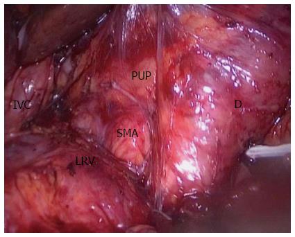
Figure 1 Superior mesentery artery was exposed from the right posterior side after complete kocherization.
D: Duodenum; IVC: Inferior vena cava; LRV: Left renal vein; PUP: Pancreatic uncinate process; SMA: Superior mesentery artery.
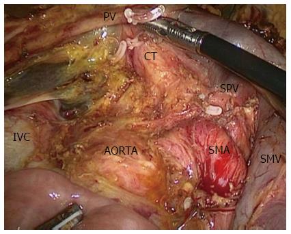
Figure 2 Local vision after removal of the specimen.
CT: Celiac trunk; IVC: Inferior vena cava; PV: Portal vein; SMA: Superior mesenteric artery; SMV: Superior mesenteric vein; SPV: Splenic vein.
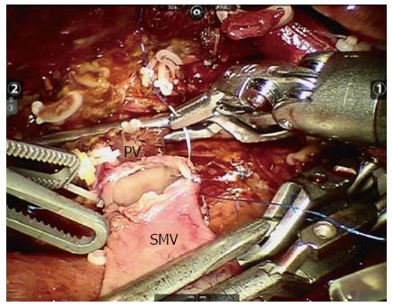
Figure 3 Vein reconstruction via robotic system.
PV: Portal vein; SMV: Superior mesenteric vein.
- Citation: Zhang YH, Zhang CW, Hu ZM, Hong DF. Pancreatic cancer: Open or minimally invasive surgery? World J Gastroenterol 2016; 22(32): 7301-7310
- URL: https://www.wjgnet.com/1007-9327/full/v22/i32/7301.htm
- DOI: https://dx.doi.org/10.3748/wjg.v22.i32.7301


