Copyright
©The Author(s) 2016.
World J Gastroenterol. Aug 21, 2016; 22(31): 7166-7174
Published online Aug 21, 2016. doi: 10.3748/wjg.v22.i31.7166
Published online Aug 21, 2016. doi: 10.3748/wjg.v22.i31.7166
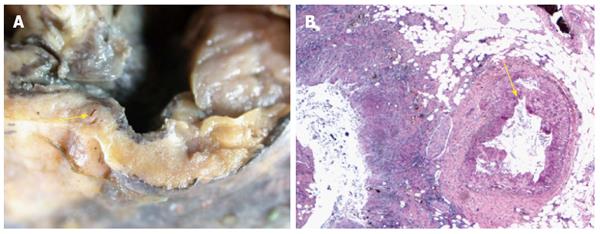
Figure 1 Post mortem examination showed large well-circumscribed duodenal diverticulum with ulcerated mucosa adjacent to eroded artery in posterior wall of first part of duodenum.
A: Artery adjacent to the ulcerated mucosa in duodenal diverticulum; B: Duodenal mucosa - Artery adjacent to the ulcerated mucosa in duodenal diverticulum (Hematoxylin and eosin × 200).
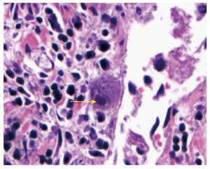
Figure 2 Owl’s eye inclusion body in duodenal diverticulum.
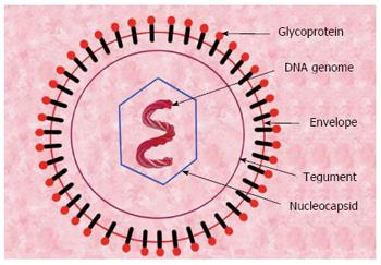
Figure 3 Cytomegalovirus structure.
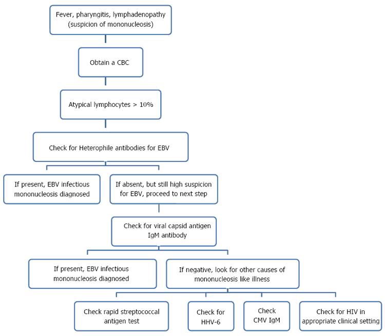
Figure 4 Approach to mononucleosis.
CMV: Cytomegalovirus.
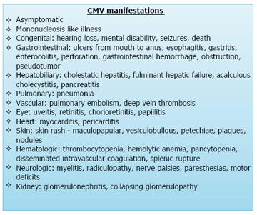
Figure 5 Manifestations of cytomegalovirus.
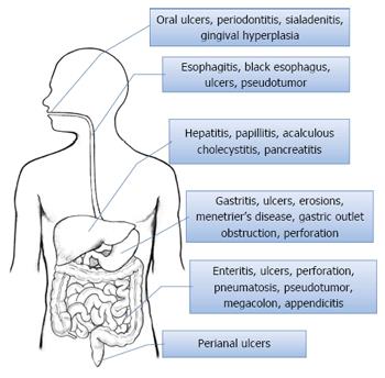
Figure 6 Cytomegalovirus gastrointestinal manifestations.
- Citation: Makker J, Bajantri B, Sakam S, Chilimuri S. Cytomegalovirus related fatal duodenal diverticular bleeding: Case report and literature review. World J Gastroenterol 2016; 22(31): 7166-7174
- URL: https://www.wjgnet.com/1007-9327/full/v22/i31/7166.htm
- DOI: https://dx.doi.org/10.3748/wjg.v22.i31.7166


