Copyright
©The Author(s) 2016.
World J Gastroenterol. Aug 14, 2016; 22(30): 6764-6775
Published online Aug 14, 2016. doi: 10.3748/wjg.v22.i30.6764
Published online Aug 14, 2016. doi: 10.3748/wjg.v22.i30.6764
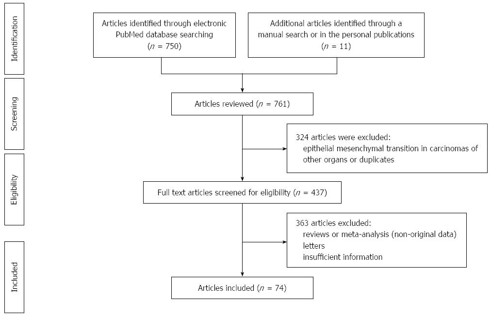
Figure 1 Preferred reported items for systematic reviews and meta-analyses (PRISMA) flow diagram adapted for data about epithelial mesenchymal transition in colorectal cancer in the PubMed database between 1995 and 2016 (First of March).
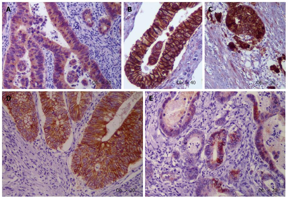
Figure 2 Epithelial mesenchymal transition-related immunoprofile of buds of colorectal cancer.
β-catenin displays cytoplasmic (A) or membrane positivity in the tumor core (B) with nuclear switch in buds (C), whereas membrane E-cadherin expression (D) is lost in the invasion front (E).
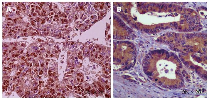
Figure 3 Nuclear positivity for Slug (A), and cytoplasmic expression of vascular endothelial growth factor-A (B) in the colorectal cancer cells.
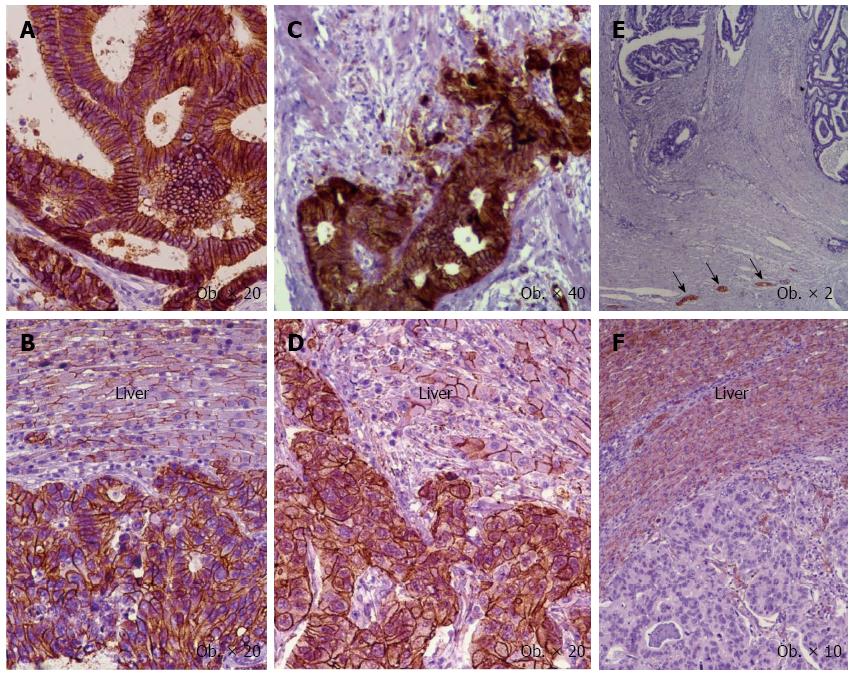
Figure 4 Immunoexpression of the epithelial mesenchymal transition-related markers in primary tumor (A, C, E) vs hepatic metastases (B, D, F).
Membrane positivity for E-cadherin (A, B) and β-catenin (C, D), and negativity for N-cadherin (E, F) with positive neural structures (arrows). The normal hepatocytes express E-cadherin (B), β-catenin (D), and N-cadherin (F).
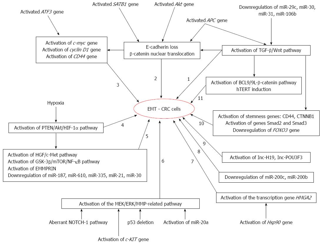
Figure 5 Molecular pathways of epithelial mesenchymal transition in colorectal cancer cells.
EMT: Epithelial mesenchymal transition; CRC: Colorectal cancer; hTERT: Human telomerase reverse transcriptase; TGF-β: Transforming growth factor-beta.
- Citation: Gurzu S, Silveanu C, Fetyko A, Butiurca V, Kovacs Z, Jung I. Systematic review of the old and new concepts in the epithelial-mesenchymal transition of colorectal cancer. World J Gastroenterol 2016; 22(30): 6764-6775
- URL: https://www.wjgnet.com/1007-9327/full/v22/i30/6764.htm
- DOI: https://dx.doi.org/10.3748/wjg.v22.i30.6764


