Copyright
©The Author(s) 2016.
World J Gastroenterol. Jul 28, 2016; 22(28): 6559-6564
Published online Jul 28, 2016. doi: 10.3748/wjg.v22.i28.6559
Published online Jul 28, 2016. doi: 10.3748/wjg.v22.i28.6559
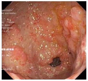
Figure 1 Gastric antral vascular ectasia observed on upper gastrointestinal endoscopy.
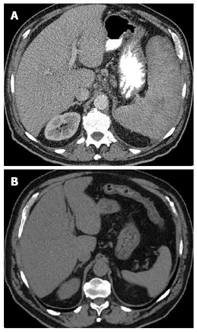
Figure 2 Images on abdominal computed tomography scan.
A: Homogeneous hepatosplenomegaly; B: Normal spleen on computed tomography scan performed two years earlier.
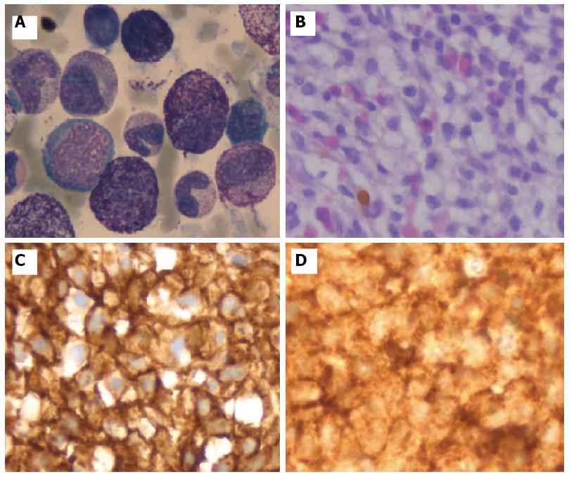
Figure 3 Immunohistochemistry study identified positive staining for CD117 and CD25.
A: Atypical spindle-shaped MC on bone marrow aspirate (HE × 100); B: Hypercellularity due to infiltration by atypical MC on bone biopsy (HE × 100); C: MC were strongly positive for CD25; and D: CD117 (D) (IHC × 100).
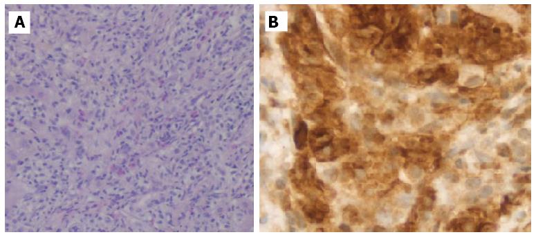
Figure 4 Microscopic images of the liver.
A: Liver biopsy showed an increased number of spindled MC (HE × 40); B: The MC are highlighted by positive CD117 staining (IHC × 100)
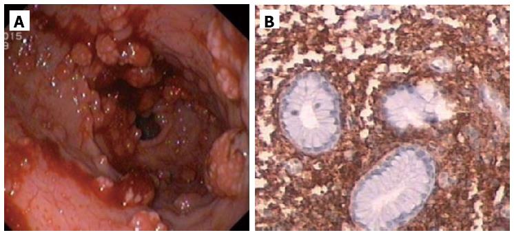
Figure 5 Extensive involvement of the stomach.
A: Endoscopic image of the stomach showed diffuse polypoid lesions with active bleeding; B: Microscopic appearance of the gastric mucosa showed infiltration by atypical MC strongly positive for CD117 (IHC × 100).
- Citation: Martins C, Teixeira C, Ribeiro S, Trabulo D, Cardoso C, Mangualde J, Freire R, Gamito &, Alves AL, Cremers I, Alves C, Neves A, Oliveira AP. Systemic mastocytosis: A rare cause of non-cirrhotic portal hypertension. World J Gastroenterol 2016; 22(28): 6559-6564
- URL: https://www.wjgnet.com/1007-9327/full/v22/i28/6559.htm
- DOI: https://dx.doi.org/10.3748/wjg.v22.i28.6559


