Copyright
©The Author(s) 2016.
World J Gastroenterol. Jul 14, 2016; 22(26): 5867-5878
Published online Jul 14, 2016. doi: 10.3748/wjg.v22.i26.5867
Published online Jul 14, 2016. doi: 10.3748/wjg.v22.i26.5867
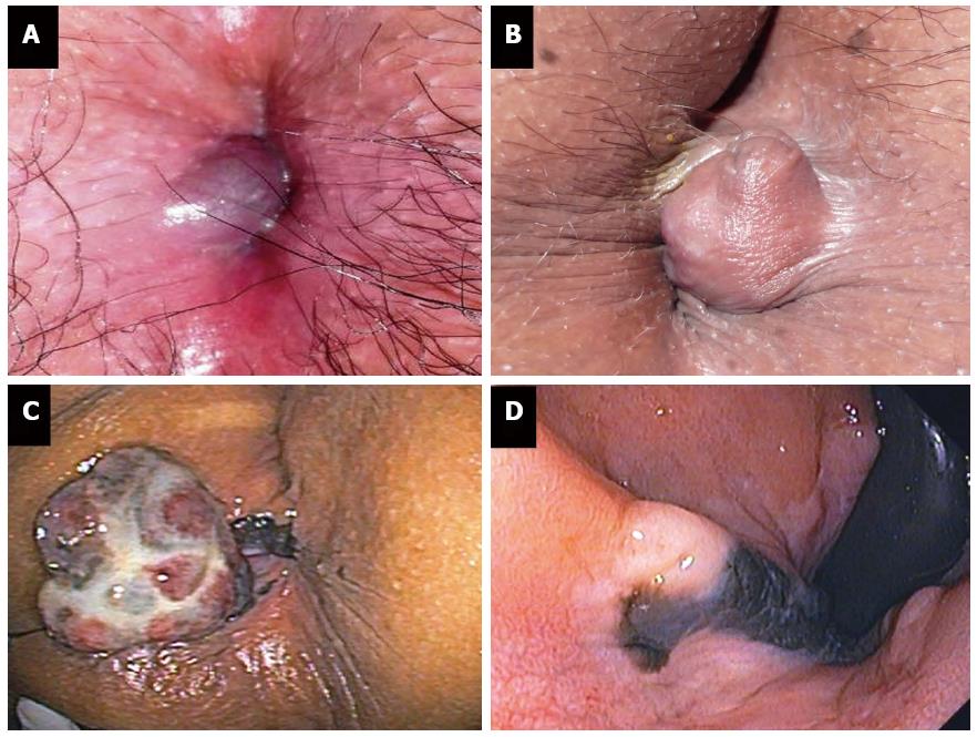
Figure 1 Acutely thrombosed external hemorrhoid (A, B) and anal melanoma (C, D).
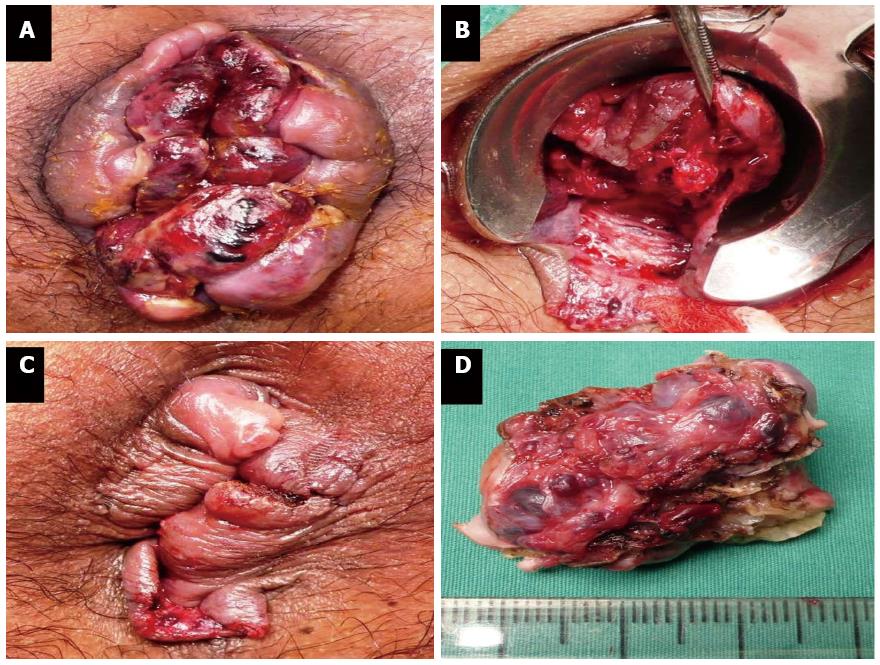
Figure 2 Urgent hemorrhoidectomy for thrombosed internal hemorrhoids (A-D).
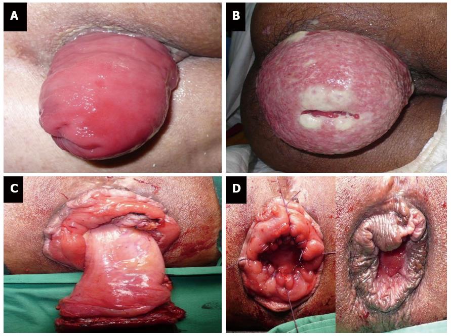
Figure 3 Rectal prolapse (A), strangulated rectal prolapse (B), and perineal rectosigmoidectomy or Altemeier’s procedure (C, D).
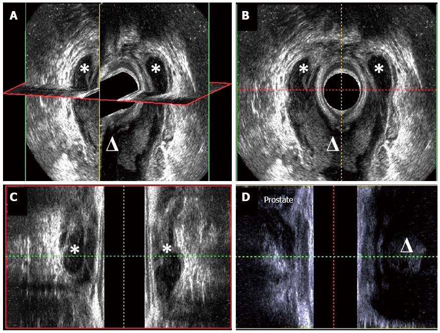
Figure 4 Three-dimensional endoanal ultrasonography of horseshoe abscess (A), cross-sectional view (B), coronal view (C) and sagittal view (D).
The asterisk means abscess in ischiorectal space and the triangle means abscess in deep postanal space.
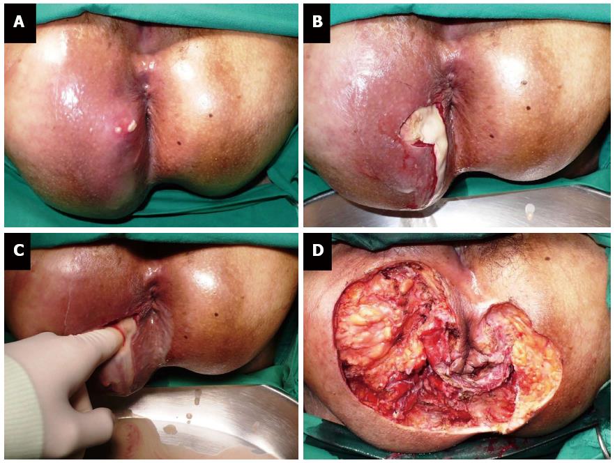
Figure 5 Perineal necrotizing fasciitis (Fournier gangrene) (A-D).
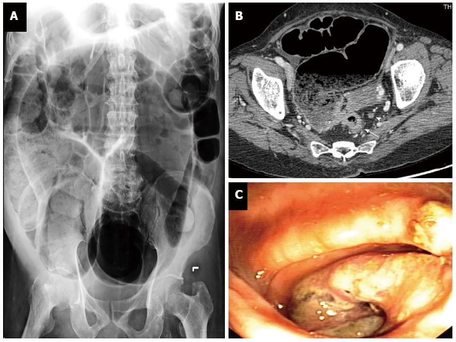
Figure 6 Obstructing rectal cancer: plain abdominal radiography (A), computerized tomography (B), and endoscopic view (C).
T3 rectal cancer (white arrow) and perirectal lymph node (arrow head).
- Citation: Lohsiriwat V. Anorectal emergencies. World J Gastroenterol 2016; 22(26): 5867-5878
- URL: https://www.wjgnet.com/1007-9327/full/v22/i26/5867.htm
- DOI: https://dx.doi.org/10.3748/wjg.v22.i26.5867


