Copyright
©The Author(s) 2016.
World J Gastroenterol. Jun 14, 2016; 22(22): 5297-5300
Published online Jun 14, 2016. doi: 10.3748/wjg.v22.i22.5297
Published online Jun 14, 2016. doi: 10.3748/wjg.v22.i22.5297
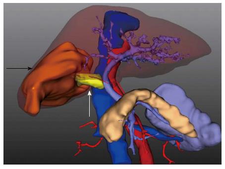
Figure 1 3D reconstruction of the tumor and portal vein tumor thrombus.
A 7-cm hepatocellular carcinoma (white arrow) was located in segments V, VI and VII. The tumor thrombus (black arrow) extended into the right branch and was adjacent to the conjunction of the portal vein.
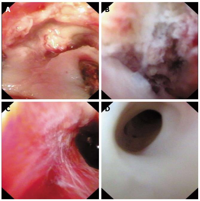
Figure 2 Endoscopic images of portal vein before and after thrombectomy.
A: Before thrombectomy, endoscopy revealed scattered tissue of tumor thrombus near the opening stump; B: Residual tumor thrombus was adhered to the inner wall of the portal vein near the conjunction; C: After repeated retraction of the residual tumor thrombus, endoscopy revealed a clean inner wall of the portal vein with no macroscopic thrombus remaining; D: The left secondary branch of the portal vein was clean with no scattered thrombus.
- Citation: Li N, Wei XB, Cheng SQ. Application of cystoscope in surgical treatment of hepatocellular carcinoma with portal vein tumor thrombus. World J Gastroenterol 2016; 22(22): 5297-5300
- URL: https://www.wjgnet.com/1007-9327/full/v22/i22/5297.htm
- DOI: https://dx.doi.org/10.3748/wjg.v22.i22.5297


