Copyright
©The Author(s) 2016.
World J Gastroenterol. Jan 14, 2016; 22(2): 718-726
Published online Jan 14, 2016. doi: 10.3748/wjg.v22.i2.718
Published online Jan 14, 2016. doi: 10.3748/wjg.v22.i2.718
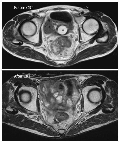
Figure 1 T2-weighted axial image of lateral pelvic lymph nodes.
A 71-year-old male experienced lateral pelvic lymph nodes (LPLN) swelling in the left internal iliac region before chemoradiotherapy (CRT). The LPLN responded to CRT, but the patient underwent laparoscopic total mesorectal excision with left LPLN dissection. Pathologic examination of LPLNs showed two left internal iliac lymph node metastases.
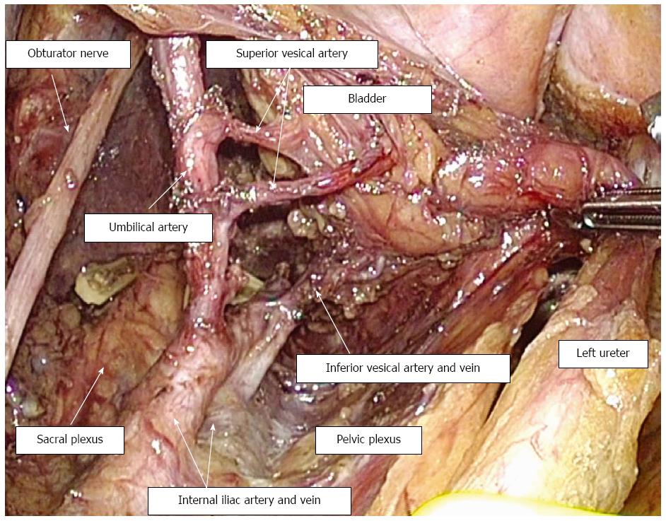
Figure 2 Surgical view after laparoscopic left lateral pelvic lymph node dissection preserving the superior and inferior vesical arteries.
The most frequent metastatic site of lateral pelvic lymph nodes was deep and around the inferior vesical vessels.
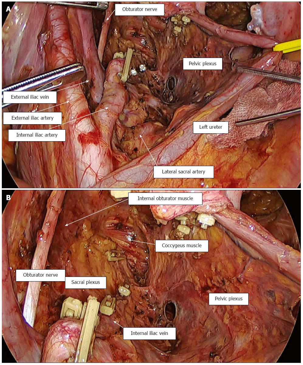
Figure 3 Surgical view after laparoscopic left lateral pelvic lymph node dissection with en bloc resection of the internal iliac artery.
A: Distant view; B: Close-up view.
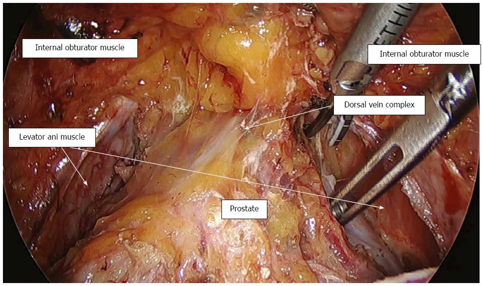
Figure 4 Surgical view after exposure of the dorsal vein complex.
This patient underwent laparoscopic total pelvic exenteration for locally recurrent rectal cancer.
- Citation: Akiyoshi T. Technical feasibility of laparoscopic extended surgery beyond total mesorectal excision for primary or recurrent rectal cancer. World J Gastroenterol 2016; 22(2): 718-726
- URL: https://www.wjgnet.com/1007-9327/full/v22/i2/718.htm
- DOI: https://dx.doi.org/10.3748/wjg.v22.i2.718


