Copyright
©The Author(s) 2016.
World J Gastroenterol. Jan 14, 2016; 22(2): 668-680
Published online Jan 14, 2016. doi: 10.3748/wjg.v22.i2.668
Published online Jan 14, 2016. doi: 10.3748/wjg.v22.i2.668
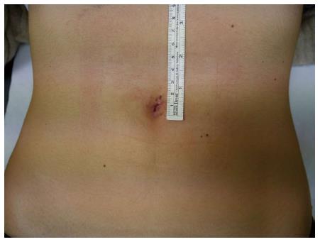
Figure 1 Single-incision laparoscopic surgery appendectomy in a 14 year old female: Umbelical scar after 8 post-operative days.
Provided by personal courtesy of Dr. S. Di Saverio, MD, FACS, FRCS.
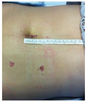
Figure 2 Single-incision laparoscopic surgery to multiport laparoscopic conversion.
Right colectomy for intended appendectomy: functional and aesthetic outcome. The procedure began as an intended single-incision laparoscopic surgery appendectomy. After discovering a wide perforation of the gangrenous cecum, the procedure was converted to a multiport laparoscopic right colectomy. With an umbelical port for the camera, two further trocars were inserted: a 5 mm trocar in the left iliac fossa and a 12 mm trocar in the left flank as an opertive port and for the insertion of the endostapler. Provided by personal courtesy of Dr. S. Di Saverio, MD, FACS, FRCS.
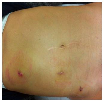
Figure 3 Laparoscopic adhesiolysis for small bowel obstruction following pedriatic surgery with median laparotomy: Functional and aesthetic outcome.
The camera was in the Hasson trocar in the paraumbelical port. Two operative 5 mm trocars were placed in the left hypocondrium and in the left iliac fossa respectively. Provided by personal courtesy of Dr. S. Di Saverio, MD, FACS, FRCS.
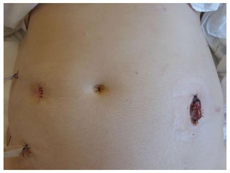
Figure 4 Laparoscopic Hartmann’s procedure for perforated Hinchey III diverticulitis in a 39 years old female: End colostomy and drains.
On the right iliac fossa a large 12 mm port was used for introducing the endostapler and for distal colonic resection. On the left flank, a fourth port was inserted for the assistant surgeon. The sigmoid was then extracted from the left flank by enlarging the port to a 4 cm incision. The end colostomy was then fashioned on the left flank using the same incision. Provided by personal courtesy of Dr. S. Di Saverio, MD, FACS, FRCS.
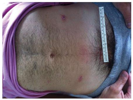
Figure 5 Resection with primary anastomosis for Hinchey IV diverticulis in a young patient: Functional and aesthetic outcome.
A suprapubic 12 mm trocar was used for introducing the endostapler and performing the fully intracorporeal anastomosis. The specimen was then extracted by enlarging the suprapubic incision to a 4 cm minilaparotomy. Provided by personal courtesy of Dr. S. Di Saverio, MD, FACS, FRCS.
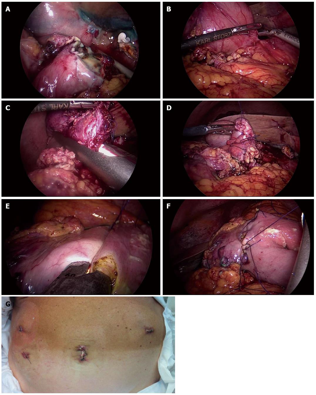
Figure 6 A four-trocar laparoscopic gastroduodenal resection for duodenal perforation.
A: “Open-book” duodenal perforation; B: Dissecting the inflamed pylorus from the head of the pancreas; C: Duodenal resection; D: Oversewing the duodenal stump; E: Performing latero-lateral intra-corporeal stapled anastomosis on the posterior stomach wall; F: Closing the enterotomy with interrupted stitches; G: Functional and aesthetic outcome. Provided by personal courtesy of Dr. S. Di Saverio, MD, FACS, FRCS.
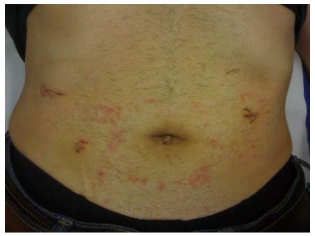
Figure 7 Nine post-operative days after laparoscopic repair of a perforated peptic ulcer: functional and aesthetic outcome.
Provided by personal courtesy of Dr. S. Di Saverio, MD, FACS, FRCS.
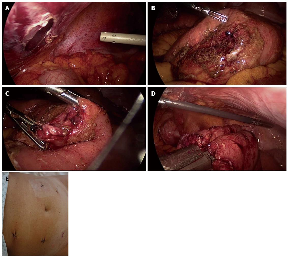
Figure 8 A large completely perforated jejunum mesenteric border with a 180° laceration of the wall.
A: Peritoneal penetration; B: Laparoscopic exploration and individuation of the site of the perforation; C: Wide perforation of the ileus; D: Performing latero-lateral intra-corporeal stapled anastomosis after a limited jejunal resection; E: Functional and aesthetic outcome. Provided by personal courtesy of Dr. S. Di Saverio, MD, FACS, FRCS.
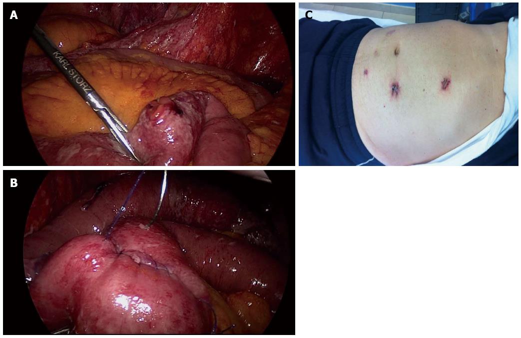
Figure 9 Laparoscopic repair of intestine perforation following a blunt abdominal trauma.
A: Individuation of the site of perforation; B: Defect repair by direct suture; C: Functional and aesthetic outcome. Provided by personal courtesy of Dr. S. Di Saverio, MD, FACS, FRCS.
- Citation: Mandrioli M, Inaba K, Piccinini A, Biscardi A, Sartelli M, Agresta F, Catena F, Cirocchi R, Jovine E, Tugnoli G, Di Saverio S. Advances in laparoscopy for acute care surgery and trauma. World J Gastroenterol 2016; 22(2): 668-680
- URL: https://www.wjgnet.com/1007-9327/full/v22/i2/668.htm
- DOI: https://dx.doi.org/10.3748/wjg.v22.i2.668


