Copyright
©The Author(s) 2016.
World J Gastroenterol. Jan 14, 2016; 22(2): 628-640
Published online Jan 14, 2016. doi: 10.3748/wjg.v22.i2.628
Published online Jan 14, 2016. doi: 10.3748/wjg.v22.i2.628
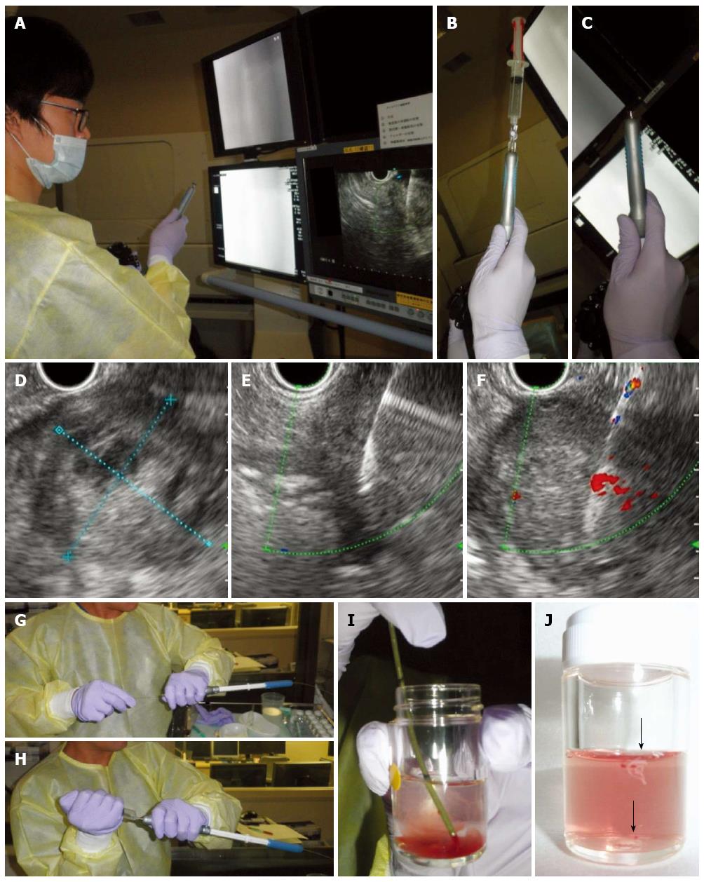
Figure 1 Actual condition of an endosonographer performing endoscopic ultrasonography guided-fine needle aspiration of a solid pancreatic lesion.
A: An endosonographer puncturing a pancreatic mass by fine needle aspiration (FNA) needle with a stylet inside the needle; B: An FNA needle aspirated with a 10 mL syringe attached to the top (Suction method); C: An FNA needle without no suction applied by a syringe (Non-suction method); D: A target pancreatic lesion, with a central necrotic area, depicted by ultrasonography and measured for its size; E: A pass at the upper side of the tumor, avoiding the central necrotic area; F: A pass at the lower part of the tumor by the fanning method; G: Expulsion of the aspirated material by insertion of a stylet; H: Flushing out of the residual material with air (This process can often be skipped.); I: A bloody sample extruded into a medium; J: Whitish components separated into another container.
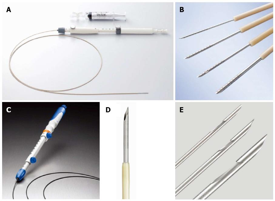
Figure 2 Variations in the fine needle aspiration needles.
A: EZ shot 2 (Olympus); B: Different size and shape of needles (EZ shot 2, from top to the bottom: 25 gauge (G), 22G, 22G with a side port); C: ExpectTM Slimline (Boston Scientific); D: 19G-flex needle of ExpectTM; E: Different size of the needles (ProCore, COOK, from top to the bottom: 25G, 22G, 19G).
- Citation: Matsubayashi H, Matsui T, Yabuuchi Y, Imai K, Tanaka M, Kakushima N, Sasaki K, Ono H. Endoscopic ultrasonography guided-fine needle aspiration for the diagnosis of solid pancreaticobiliary lesions: Clinical aspects to improve the diagnosis. World J Gastroenterol 2016; 22(2): 628-640
- URL: https://www.wjgnet.com/1007-9327/full/v22/i2/628.htm
- DOI: https://dx.doi.org/10.3748/wjg.v22.i2.628


