Copyright
©The Author(s) 2016.
World J Gastroenterol. May 14, 2016; 22(18): 4610-4614
Published online May 14, 2016. doi: 10.3748/wjg.v22.i18.4610
Published online May 14, 2016. doi: 10.3748/wjg.v22.i18.4610
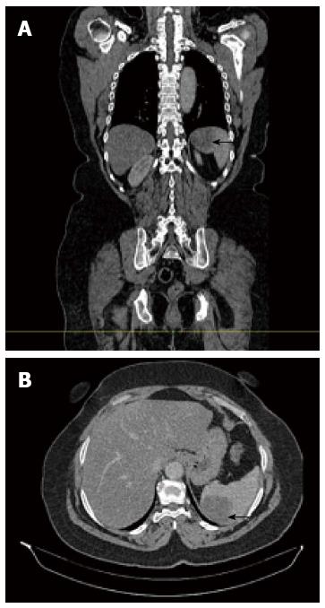
Figure 1 Abdominal computed scan demonstrating, in sagittal (A) and axial (B) views, a single low-density lesion of the spleen which measures 4.
9 cm in diameter.
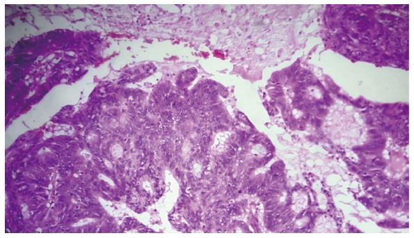
Figure 2 Adenocarcinoma spread the splenic parenchyma (hematoxylin-eosin staining, magnification × 20).
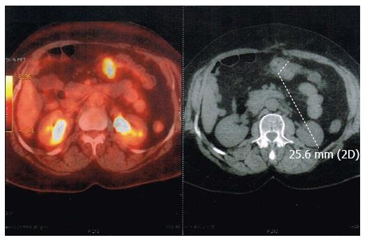
Figure 3 Fluorodeoxyglucose-positron emission tomography with high metabolic activity in the abdominal cavity.
- Citation: Abdou J, Omor Y, Boutayeb S, Elkhannoussi B, Errihani H. Isolated splenic metastasis from colon cancer: Case report. World J Gastroenterol 2016; 22(18): 4610-4614
- URL: https://www.wjgnet.com/1007-9327/full/v22/i18/4610.htm
- DOI: https://dx.doi.org/10.3748/wjg.v22.i18.4610


