Copyright
©The Author(s) 2016.
World J Gastroenterol. May 14, 2016; 22(18): 4515-4528
Published online May 14, 2016. doi: 10.3748/wjg.v22.i18.4515
Published online May 14, 2016. doi: 10.3748/wjg.v22.i18.4515
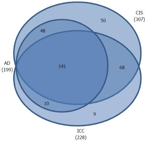
Figure 1 Venn diagrams of comparisons of the differentially expressed proteins from the different stages compared to the NC group.
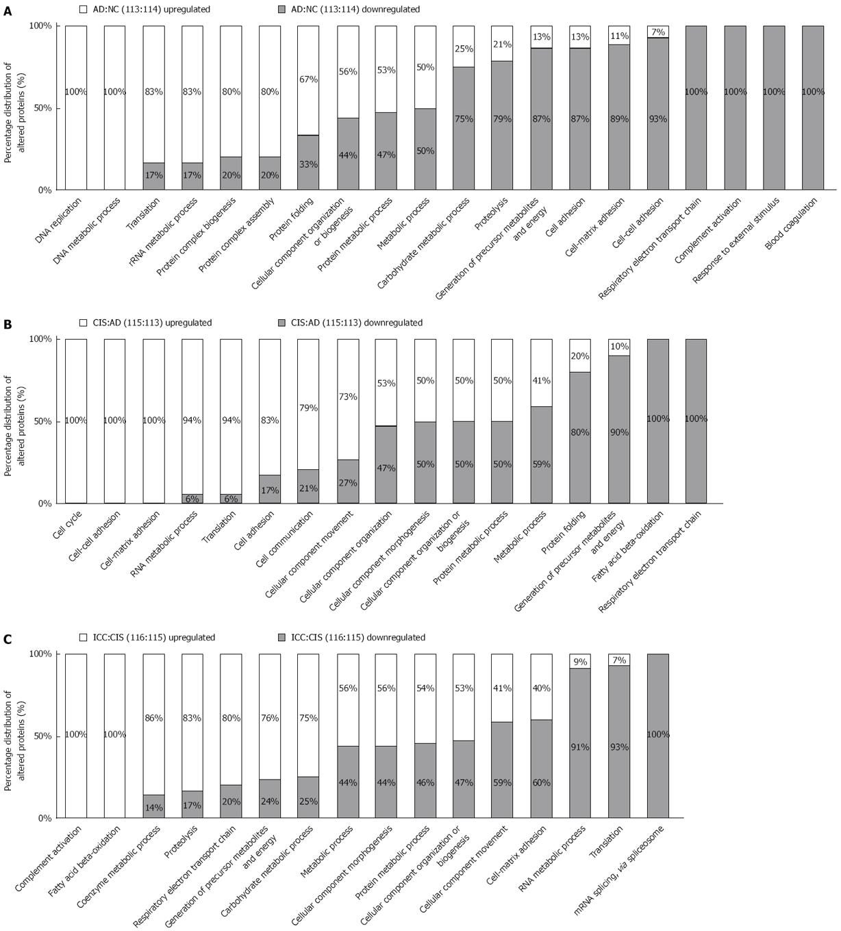
Figure 2 Functional distribution of differentially expressed proteins between adjacent stages of the colon carcinogenesis process.
Functional classification of differentially expressed proteins from AD vs NC, CIS vs AD and ICC vs CIS were assigned to “biological process” subcategories. Only significant subcategories for “biological process” are presented. Each subcategory is presented as the percentage of up- and down-regulated proteins.
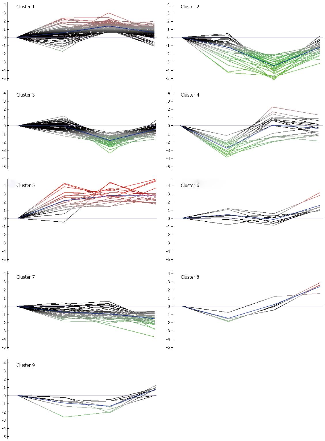
Figure 3 K-mean clusters of differentially expressed proteins.
These proteins could be clustered into nine clusters. According to the average tendency, the nine clusters can be arbitrarily categorized into three groups. Group 1 includes clusters 1 and 5, in which the abundance of proteins progressively increased during the colon carcinogenic process. Group 2 consists of clusters 2 and 7, in which the abundance of proteins progressively decreased during the process. Group 3 consists of clusters 3, 4, 6, 8 and 9, in which the abundance of proteins was significantly up-regulated or down-regulated in certain stages of the process.
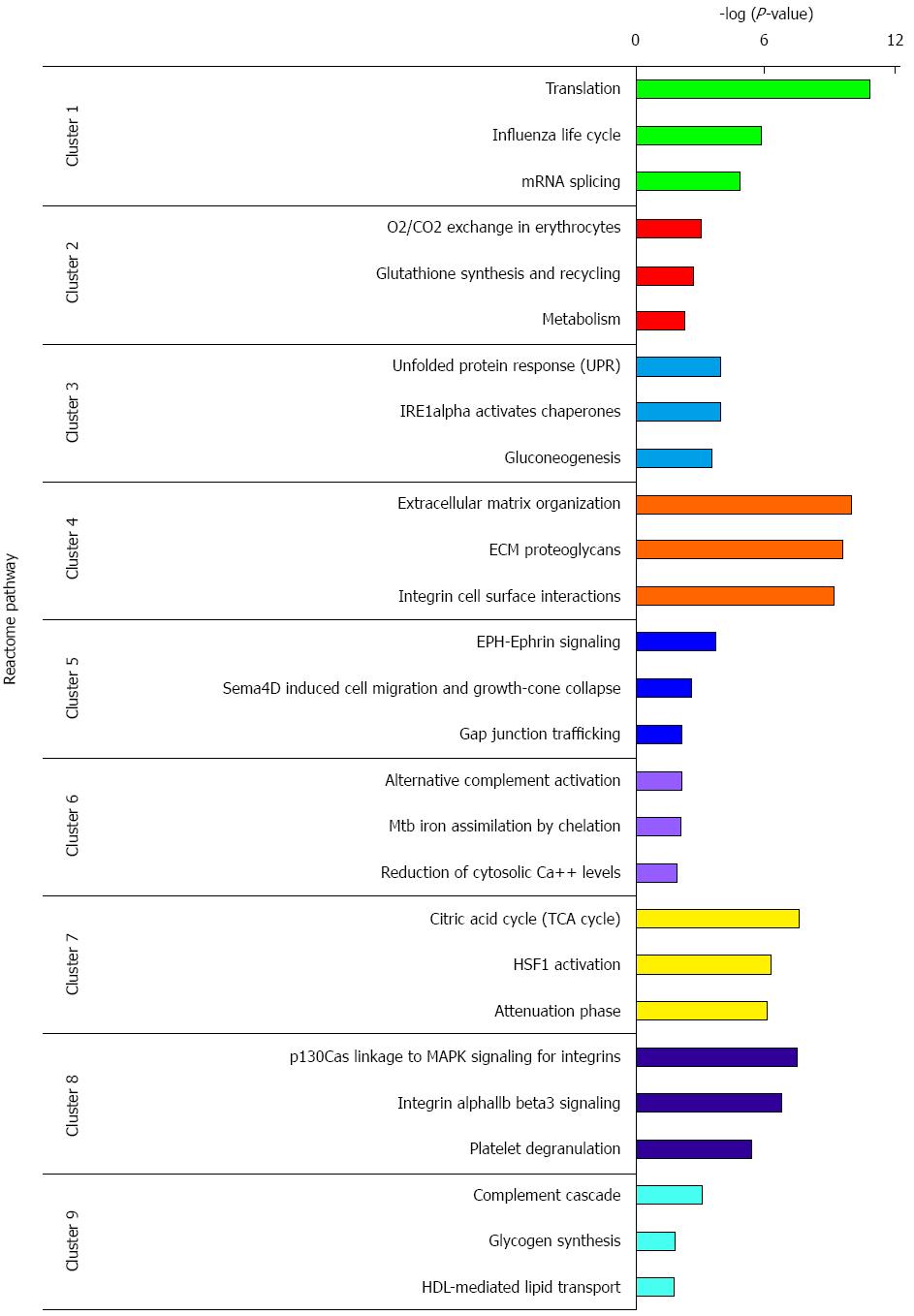
Figure 4 REACTOME pathway analysis of differential expressed proteins in each cluster.
Only top 3 significant pathways are presented. Each pathway is presented as negative logarithm of P-value.
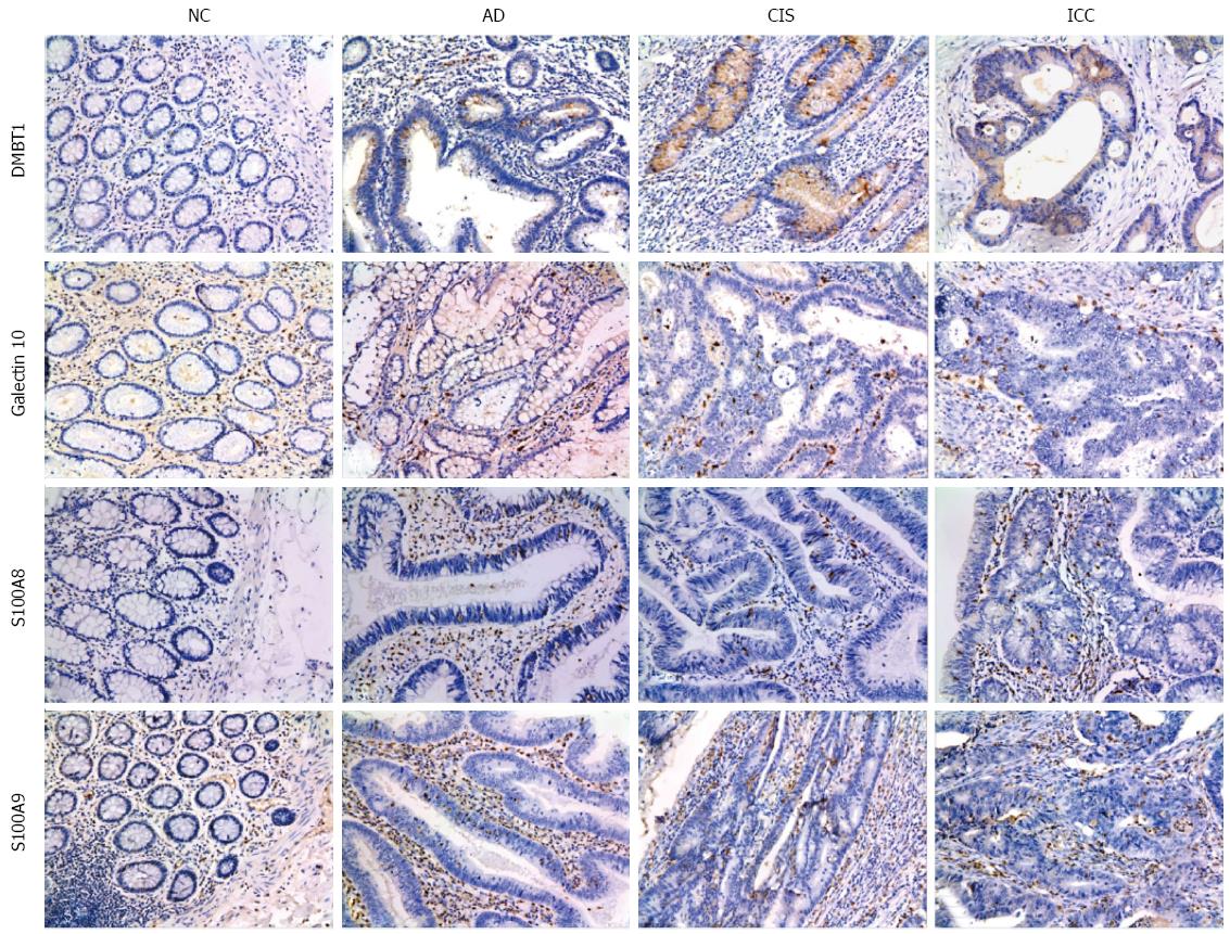
Figure 5 Representative results of immunohistochemistry show the expression of DMBT1, S100A9, Galectin-10 and S100A8 in the NC, AD, CIS and ICC (Original magnification, × 200).
DMBT1 immunostaining in NC, AD, CIS and ICC. Negative staining was observed in NC, moderate in AD tissues, and strong cytoplasmic staining in CIS and ICC tissues. S100A9 immunostaining in NC, AD, CIS and ICC. Negative staining was found in NC, weak intralesional staining in AD, moderate intralesional staining in CIS and strong intralesional staining in ICC. Galectin-10 immunostaining in NC, AD, CIS and ICC. Negative staining was found in NC, weak staining in AD and CIS, and moderate staining in ICC. S100A8 immunostaining in NC, AD, CIS and ICC. Negative staining was observed in NC, moderate intralesional staining in AD and CIS, and strong intralesional staining in ICC.
- Citation: Peng F, Huang Y, Li MY, Li GQ, Huang HC, Guan R, Chen ZC, Liang SP, Chen YH. Dissecting characteristics and dynamics of differentially expressed proteins during multistage carcinogenesis of human colorectal cancer. World J Gastroenterol 2016; 22(18): 4515-4528
- URL: https://www.wjgnet.com/1007-9327/full/v22/i18/4515.htm
- DOI: https://dx.doi.org/10.3748/wjg.v22.i18.4515


