Copyright
©The Author(s) 2016.
World J Gastroenterol. Apr 28, 2016; 22(16): 4264-4269
Published online Apr 28, 2016. doi: 10.3748/wjg.v22.i16.4264
Published online Apr 28, 2016. doi: 10.3748/wjg.v22.i16.4264
Figure 1 Images of patient 1 who underwent urgent endoscopic ultrasound-guided choledochoduodenostomy.
A: EUS-guided common bile duct (CBD) puncture; B: EUS-guided cholangiography showed large piled-up CBD stones. Fistula track was dilated using a 4-mm balloon catheter; C: A covered metallic stent and an endoscopic nasobiliary drainage catheter were successfully placed via the duodenum bulb. EUS: Endoscopic ultrasound.
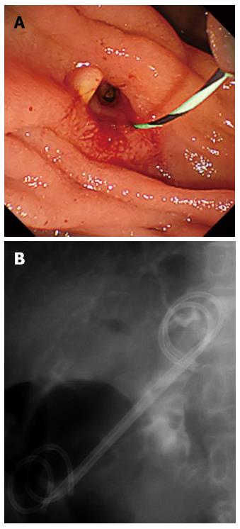
Figure 2 Images of biliary cannulation and plastic stent placement via an endoscopic ultrasound-guided choledochoduodenostomy fistula.
A: A matured fistula was created where the removed metallic stent was placed. The common bile duct (CBD) was cannulated via the EUS-CDS fistula using an ERCP catheter and a guidewire was inserted; B: Two 7-Fr double pigtail plastic stents were placed into the CBD via the EUS-CDS fistula. EUS-CDS: Endoscopic ultrasound-guided choledochoduodenostomy; ERCP: Endoscopic retrograde cholangiopancreatography.
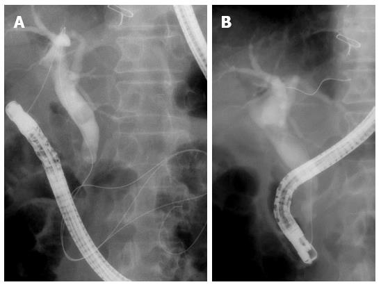
Figure 3 Images of a rendezvous technique via the endoscopic ultrasound-guided choledochoduodenostomy fistula.
A: The common bile duct was cannulated via the EUS-CDS fistula and a guidewire was advanced into the duodenum through the papilla; B: The guidewire was caught in the duodenum and transpapillary biliary cannulation was succeeded.
- Citation: Minaga K, Kitano M, Imai H, Yamao K, Kamata K, Miyata T, Omoto S, Kadosaka K, Yoshikawa T, Kudo M. Urgent endoscopic ultrasound-guided choledochoduodenostomy for acute obstructive suppurative cholangitis-induced sepsis. World J Gastroenterol 2016; 22(16): 4264-4269
- URL: https://www.wjgnet.com/1007-9327/full/v22/i16/4264.htm
- DOI: https://dx.doi.org/10.3748/wjg.v22.i16.4264


