Copyright
©The Author(s) 2016.
World J Gastroenterol. Apr 28, 2016; 22(16): 4259-4263
Published online Apr 28, 2016. doi: 10.3748/wjg.v22.i16.4259
Published online Apr 28, 2016. doi: 10.3748/wjg.v22.i16.4259
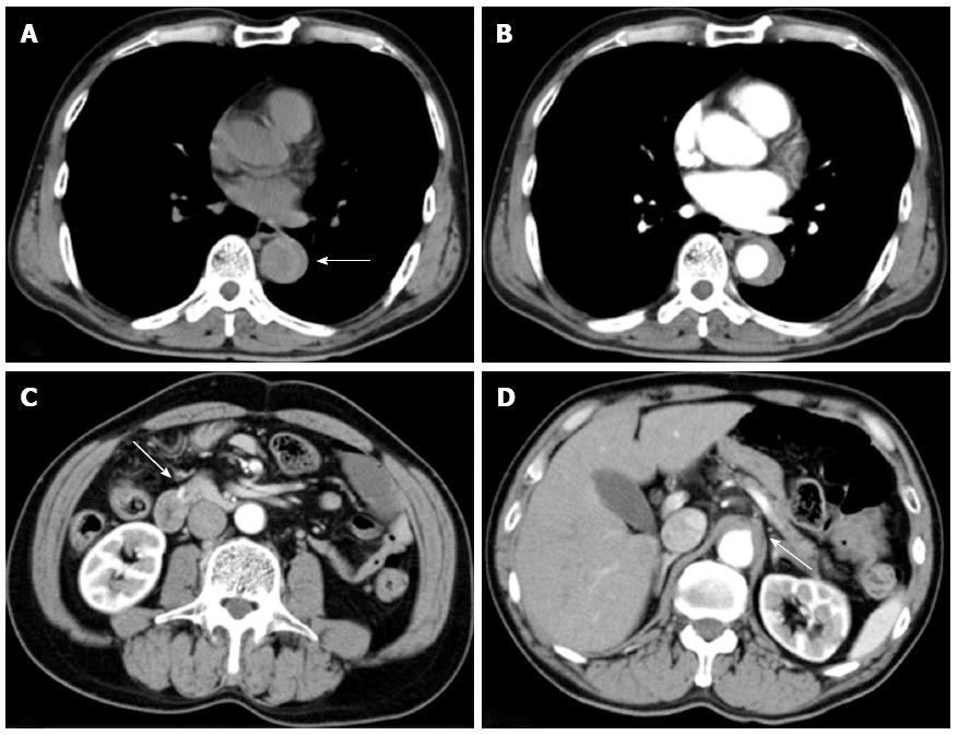
Figure 1 Initial computed tomography performed at local hospital before the onset of hypotension.
A: Plain computed tomography (CT) demonstrating dilated descending aorta and hyperattenuated collection located eccentrically within the aortic wall (arrow); B: Contrast-enhanced CT revealed crescentic and asymmetric wall thickening of the descending aorta: Contrast is not visualized within the aortic media. Intimal flap was not detected; C: An aneurysm at the anteroinferior pancreaticoduodenal artery (arrow); D: The occluded root of the coeliac artery (arrow).
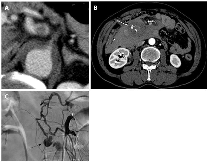
Figure 2 Subsequent contrast-enhanced computed tomography and arteriography performed at our hospital, after the onset of hypotension.
A: Multidetector computed tomography (CT) performed at our hospital showing the root of the coeliac artery compressed and occluded by the false lumen more clearly than that performed at a local hospital; B: CT scan showing a large haematoma in the right prerenal space (arrow head) and the aneurysm of anteroinferior pancreaticoduodenal artery (arrow); C: Arteriogram of the superior mesenteric artery (SMA) showing blood flow reversal from the SMA to the hepatic artery through the pancreaticoduodenal arcade, due to coeliac trunk occlusion and a saccular aneurysm of the inferior pancreaticoduodenal artery (PDA) (arrow).
- Citation: Sakatani A, Doi Y, Kitayama T, Matsuda T, Sasai Y, Nishida N, Sakamoto M, Uenoyama N, Kinoshita K. Pancreaticoduodenal artery aneurysm associated with coeliac artery occlusion from an aortic intramural hematoma. World J Gastroenterol 2016; 22(16): 4259-4263
- URL: https://www.wjgnet.com/1007-9327/full/v22/i16/4259.htm
- DOI: https://dx.doi.org/10.3748/wjg.v22.i16.4259


