Copyright
©The Author(s) 2016.
World J Gastroenterol. Apr 21, 2016; 22(15): 3945-3951
Published online Apr 21, 2016. doi: 10.3748/wjg.v22.i15.3945
Published online Apr 21, 2016. doi: 10.3748/wjg.v22.i15.3945
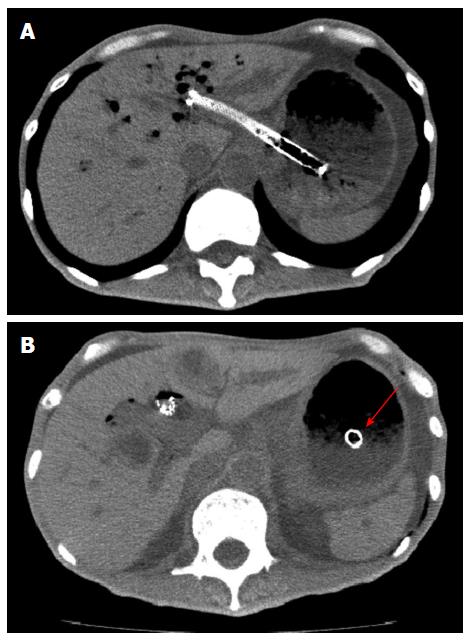
Figure 1 Dislocation of endoscopic ultrasound-guided hepaticogastrostomy metallic stent.
(A) EUS-HGS was performed for gastric cancer patient. (B) Because of tumor growth, dislocation of EUS-HGS metallic stent was seen (arrow). EUS-HGS: Endoscopic ultrasound-guided hepaticogastrostomy.
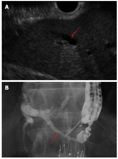
Figure 2 Technical tips of puncture on endoscopic ultrasound-guided hepaticogastrostomy.
(A) To advance the guidewire toward the hepatic hilum, the bile duct that runs from the upper left to the lower right on EUS imaging should be punctured (arrow) (B) fluoroscopic imaging (arrow). EUS-HGS: Endoscopic ultrasound-guided hepaticogastrostomy.
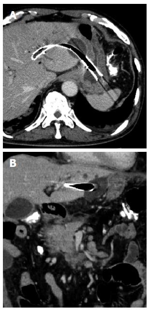
Figure 3 Biloma after endoscopic ultrasound-guided hepaticogastrostomy.
Long procedure time was needed, therefore, bile leak was increased.
- Citation: Ogura T, Higuchi K. Technical tips for endoscopic ultrasound-guided hepaticogastrostomy. World J Gastroenterol 2016; 22(15): 3945-3951
- URL: https://www.wjgnet.com/1007-9327/full/v22/i15/3945.htm
- DOI: https://dx.doi.org/10.3748/wjg.v22.i15.3945


