Copyright
©The Author(s) 2016.
World J Gastroenterol. Apr 14, 2016; 22(14): 3875-3878
Published online Apr 14, 2016. doi: 10.3748/wjg.v22.i14.3875
Published online Apr 14, 2016. doi: 10.3748/wjg.v22.i14.3875
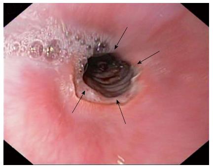
Figure 1 Upper endoscopy showing a giant ulcer of the distal esophagus (arrows).
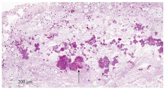
Figure 2 Histopathological examination of ulcer showed esophagitis with ulceration and underlying granulation tissue, with many bacterial colonies and fungal spores (PAS-positive) (arrow).
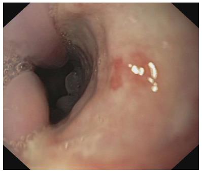
Figure 3 Endoscopy demonstrated the near complete healing of the ulcer.
- Citation: Veroux M, Aprile G, Amore FF, Corona D, Giaquinta A, Veroux P. Rare cause of odynophagia: Giant esophageal ulcer. World J Gastroenterol 2016; 22(14): 3875-3878
- URL: https://www.wjgnet.com/1007-9327/full/v22/i14/3875.htm
- DOI: https://dx.doi.org/10.3748/wjg.v22.i14.3875


