Copyright
©The Author(s) 2016.
World J Gastroenterol. Apr 14, 2016; 22(14): 3758-3768
Published online Apr 14, 2016. doi: 10.3748/wjg.v22.i14.3758
Published online Apr 14, 2016. doi: 10.3748/wjg.v22.i14.3758
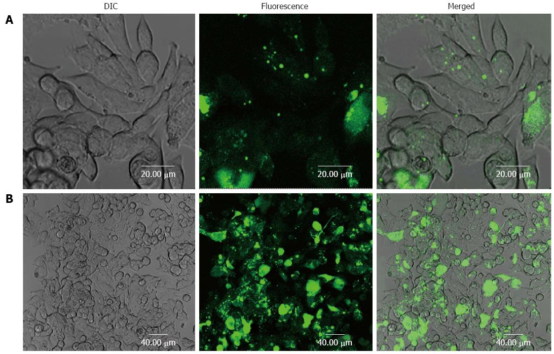
Figure 1 Confocal microscopy of LS180 cancer cells incubated with FITC-BSA-PCL and FITC-antiEGFR-BSA-PCL for 4 h.
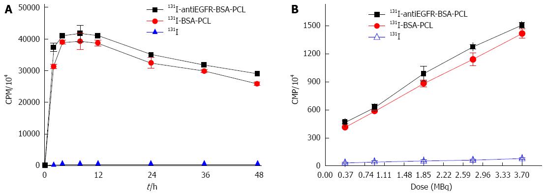
Figure 2 131I uptake in LS180 cells.
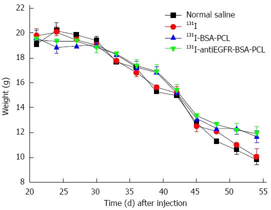
Figure 3 Changes in the animal body weight.
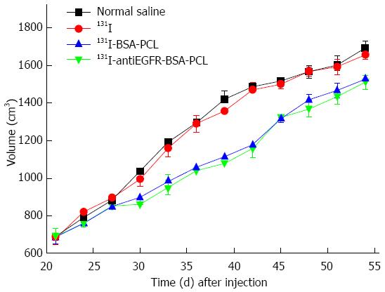
Figure 4 Changes in tumor volume.
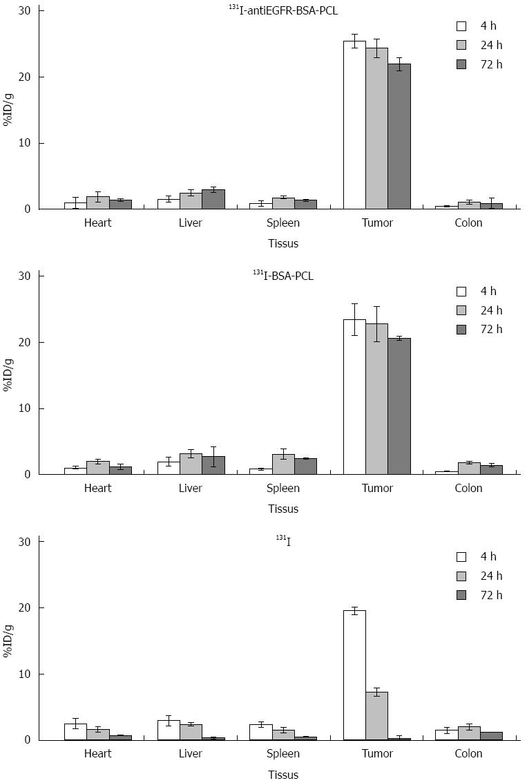
Figure 5 Tissue distribution of 131I-antiEGFR-BSA-PCL, 131I-BSA-PCL and 131I.
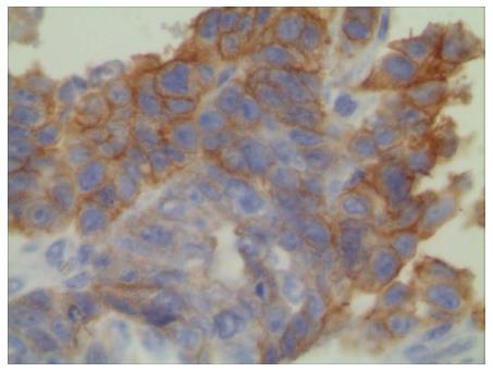
Figure 6 Immunohistochemical staining of LS180 colorectal cancer.
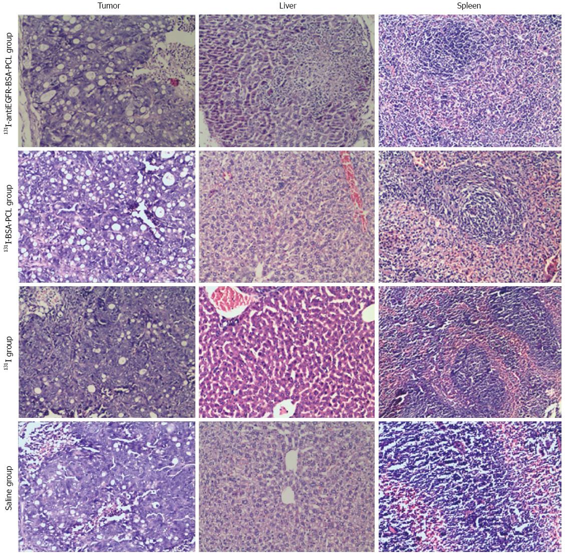
Figure 7 Histopathology of the liver, spleen, and tumor tissue at 3 d after the end of the treatment (× 100).
- Citation: Li W, Ji YH, Li CX, Liu ZY, Li N, Fang L, Chang J, Tan J. Evaluation of therapeutic effectiveness of 131I-antiEGFR-BSA-PCL in a mouse model of colorectal cancer. World J Gastroenterol 2016; 22(14): 3758-3768
- URL: https://www.wjgnet.com/1007-9327/full/v22/i14/3758.htm
- DOI: https://dx.doi.org/10.3748/wjg.v22.i14.3758


