Copyright
©The Author(s) 2016.
World J Gastroenterol. Apr 7, 2016; 22(13): 3558-3563
Published online Apr 7, 2016. doi: 10.3748/wjg.v22.i13.3558
Published online Apr 7, 2016. doi: 10.3748/wjg.v22.i13.3558
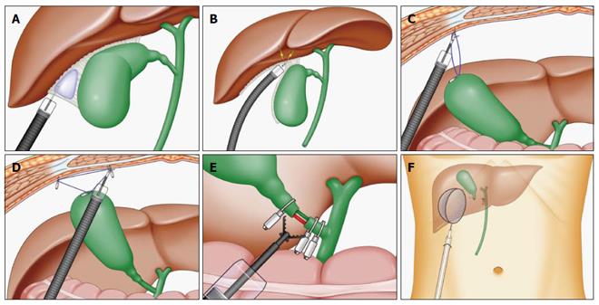
Figure 1 Conceptual images of endoscopic cholecystectomy via single port.
A: Local injection of saline to the gallbladder bed; B: Resection of the gallbladder from the liver was performed without any damage or bleeding due to sufficient space of local injection; C: A ring-shaped thread was placed, and one side of the ring was clipped to the fundus of the gallbladder and the other to the abdominal wall; D: The third clip was hooked to 1 side of the ring-shaped thread. The thread was hooked crosswise to the 2 supporting points of the gallbladder and abdominal wall; E: The neck of the gallbladder was cut using scissor forceps; F: Resected gallbladder was retrieved using endoscopic net forceps.
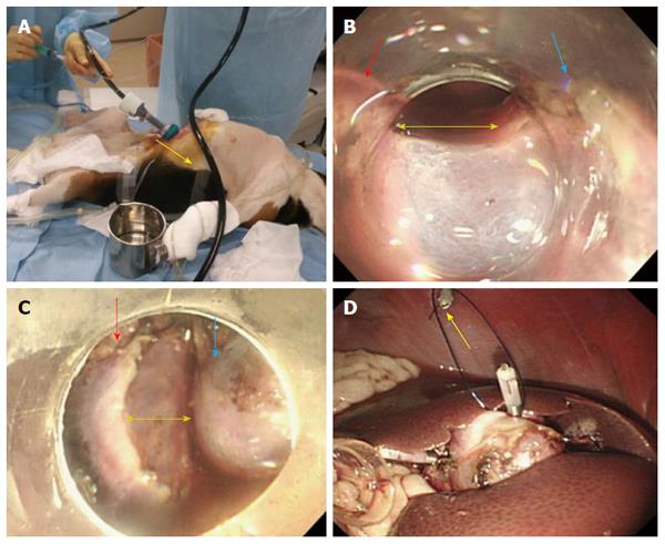
Figure 2 In vivo two dogs’ experiments.
A: Under general anesthesia, only 1 port (12 mm in diameter) was inserted using an open technique; B: A flexible endoscope with a tip attachment was inserted between the fundus of gallbladder (blue arrow) and liver (red arrow), and a layer of loose connective tissue was opened (yellow arrow); C: Complete dissection (yellow arrow) of the gallbladder bed (blue arrow) from liver (red arrow); D: One side of the ring-shaped thread was clipped to the fundus of the gallbladder and the other to the abdominal wall (yellow arrow).
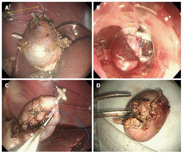
Figure 3 The ring-shaped thread technique.
A: The thread was hooked crosswise to the 2 supporting points (yellow arrow) of the gallbladder (blue arrow) and abdominal wall. Red arrow revealed the liver; B: The neck of the gallbladder was cut using scissor forceps (yellow arrow: neck of the gallbladder; blue arrows: gallbladder vein; red arrow: gallbladder artery; green arrow: cystic duct); C: The gallbladder was completely resected and put upon (blue curved arrow) the liver (red arrow); D: Resected gallbladder bed was very smooth without blood and damage.
- Citation: Mori H, Kobayashi N, Kobara H, Nishiyama N, Fujihara S, Chiyo T, Ayaki M, Nagase T, Masaki T. Novel and safer endoscopic cholecystectomy using only a flexible endoscope via single port. World J Gastroenterol 2016; 22(13): 3558-3563
- URL: https://www.wjgnet.com/1007-9327/full/v22/i13/3558.htm
- DOI: https://dx.doi.org/10.3748/wjg.v22.i13.3558


