Copyright
©The Author(s) 2016.
World J Gastroenterol. Mar 28, 2016; 22(12): 3506-3510
Published online Mar 28, 2016. doi: 10.3748/wjg.v22.i12.3506
Published online Mar 28, 2016. doi: 10.3748/wjg.v22.i12.3506
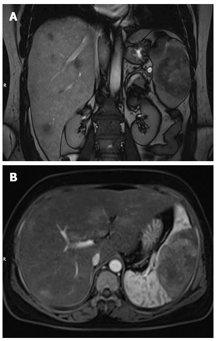
Figure 1 Magnetic resonance imaging.
A: Coronal T2-weighted image revealed multiple nodular lesions in the liver parenchyma; B: Contrast-enhanced magnetic resonance imaging showed marked heterogeneous enhancement in the splenic mass, and multiple high signal intensity lesions in the liver.
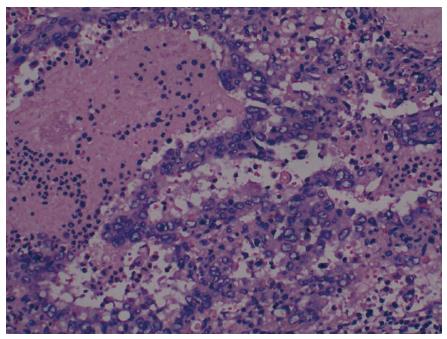
Figure 2 Histopathologic findings of the splenic specimen.
Clusters of spindle tumor cells infiltrating the spleen parenchyma (HE stain; magnification × 200).
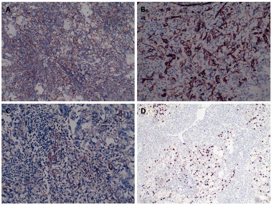
Figure 3 Immunohistochemical examination of the splenic specimen.
A: Specimen stained positive for CD31 (magnification × 200); B: Specimen stained positive for CD34 (magnification × 200); C: Specimen stained positive for FVIII (magnification × 200); D: Ki-67 proliferation index less than 30% (magnification × 100).
- Citation: Yang KF, Li Y, Wang DL, Yang JW, Wu SY, Xiao WD. Primary splenic angiosarcoma with liver metastasis: A case report and literature review. World J Gastroenterol 2016; 22(12): 3506-3510
- URL: https://www.wjgnet.com/1007-9327/full/v22/i12/3506.htm
- DOI: https://dx.doi.org/10.3748/wjg.v22.i12.3506


