Copyright
©The Author(s) 2016.
World J Gastroenterol. Mar 28, 2016; 22(12): 3381-3391
Published online Mar 28, 2016. doi: 10.3748/wjg.v22.i12.3381
Published online Mar 28, 2016. doi: 10.3748/wjg.v22.i12.3381
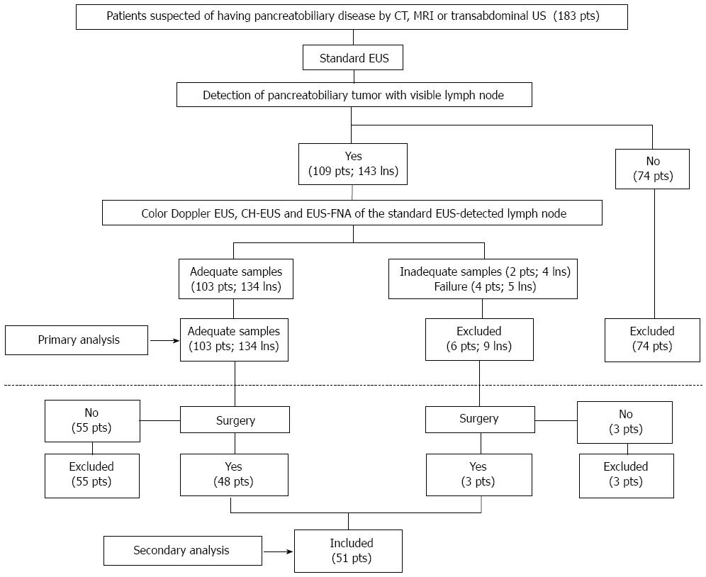
Figure 1 Schematic depiction of patient selection and exclusion criteria.
pts: Patients; lns: Lymph nodes. CT: Computed tomography; MRI: Magnetic resonance imaging; EUS: Endoscopic ultrasonography; FNA: Fine needle aspiration.
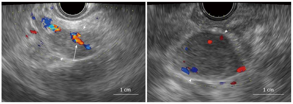
Figure 2 Typical examples of lymph nodes with (A) and without (B) a central intranodal blood vessel on color Doppler endoscopic ultrasonography.
An apparently visible lymph node was detected in both A and B (arrowheads). A shows a tubular structure that was ≥ 1 mm in diameter and was located toward the center of the lymph node and demonstrated blood flow on color Doppler endoscopic ultrasonography (arrow).
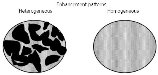
Figure 3 Contrast-enhanced harmonic endoscopic ultrasonography -determined enhancement patterns of the lymph node.
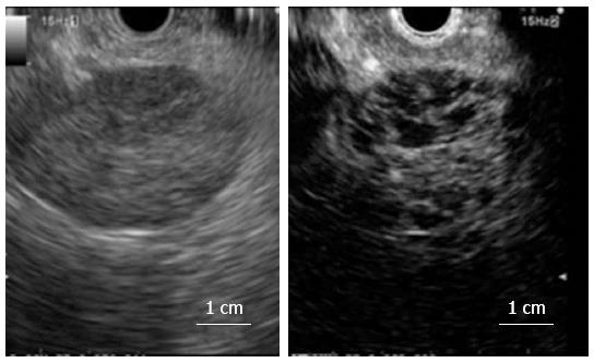
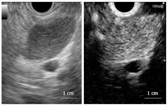
Figure 4 Typical example of a metastatic lymph node that shows heterogeneous enhancement on contrast-enhanced harmonic endoscopic ultrasonography (left, fundamental B mode; right, contrast harmonic mode).
Figure 5 Typical example of a reactive lymph node that shows homogeneous enhancement on contrast-enhanced harmonic endoscopic ultrasonography (left, fundamental B mode; right, contrast harmonic mode).
- Citation: Miyata T, Kitano M, Omoto S, Kadosaka K, Kamata K, Imai H, Sakamoto H, Nisida N, Harwani Y, Murakami T, Takeyama Y, Chiba Y, Kudo M. Contrast-enhanced harmonic endoscopic ultrasonography for assessment of lymph node metastases in pancreatobiliary carcinoma. World J Gastroenterol 2016; 22(12): 3381-3391
- URL: https://www.wjgnet.com/1007-9327/full/v22/i12/3381.htm
- DOI: https://dx.doi.org/10.3748/wjg.v22.i12.3381


