Copyright
©The Author(s) 2016.
World J Gastroenterol. Mar 14, 2016; 22(10): 3052-3055
Published online Mar 14, 2016. doi: 10.3748/wjg.v22.i10.3052
Published online Mar 14, 2016. doi: 10.3748/wjg.v22.i10.3052
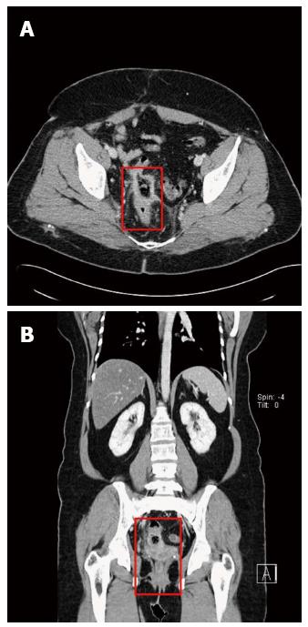
Figure 1 Computed tomography during venous phase of contrast injection.
Each red rectangle in axial (A) and coronal (B) computed tomographic images indicates wall thickening and mucosal enhancement of the rectosigmoid colon.
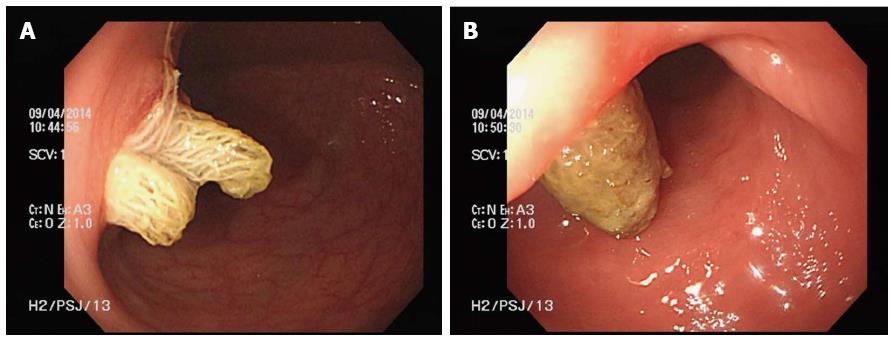
Figure 2 Colonoscopic findings.
The penetrated surgical sponge had formed two openings of the fistula to the sigmoid colon (A) and the rectum (B), respectively.
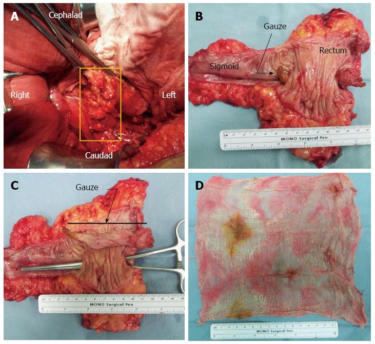
Figure 3 Operative and specimen findings.
A mass like lesion found in the left lateral wall of the rectosigmoid colon (A); Surgical sponge penetrated to the submucosal layer of the sigmoid colon and rectum and migrated to form a fistula (B and C); Retained surgical sponge (D).
- Citation: Shin WY, Im CH, Choi SK, Choe YM, Kim KR. Transmural penetration of sigmoid colon and rectum by retained surgical sponge after hysterectomy. World J Gastroenterol 2016; 22(10): 3052-3055
- URL: https://www.wjgnet.com/1007-9327/full/v22/i10/3052.htm
- DOI: https://dx.doi.org/10.3748/wjg.v22.i10.3052


