Copyright
©The Author(s) 2016.
World J Gastroenterol. Jan 7, 2016; 22(1): 112-125
Published online Jan 7, 2016. doi: 10.3748/wjg.v22.i1.112
Published online Jan 7, 2016. doi: 10.3748/wjg.v22.i1.112
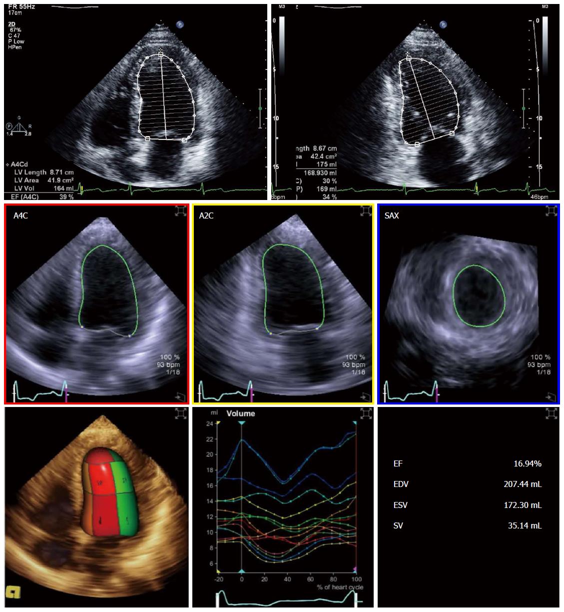
Figure 1 Ejection fraction determination using two-dimensional (biplane Simpson’s method - upper panel) and three-dimensional echocardiography (fully automated software - lower panel).
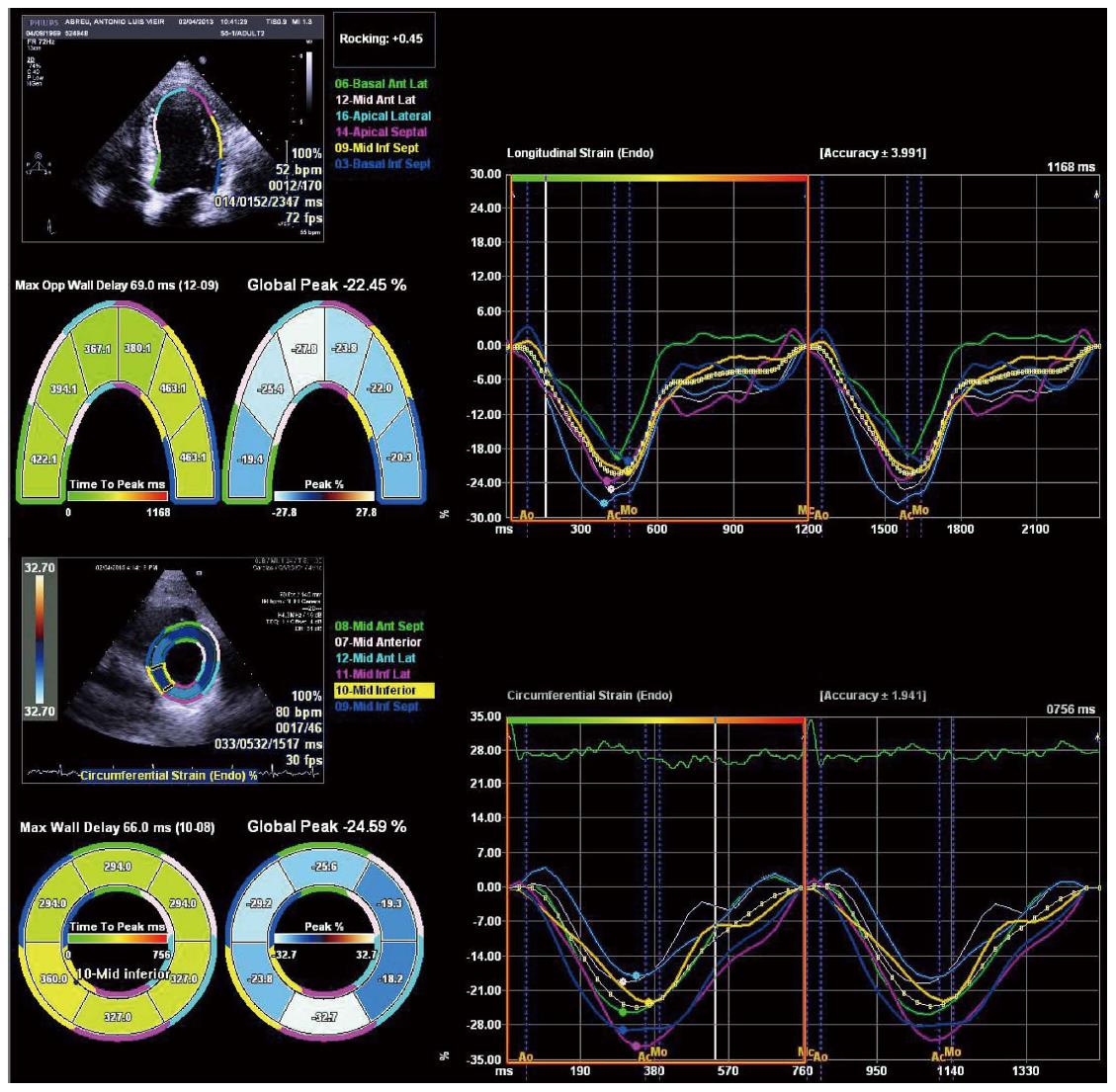
Figure 2 Left ventricular deformation analysis using speckle tracking echocardiography.
Longitudinal (top panel) and circumferential (lower panel) strain curves are displayed.
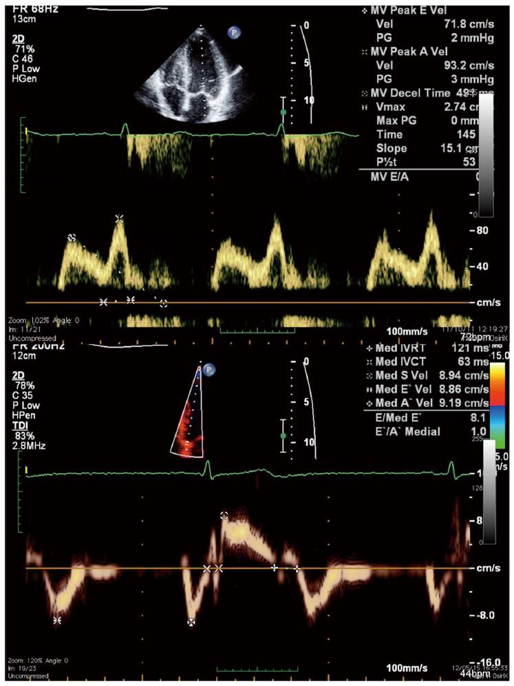
Figure 3 Mitral inflow velocities using pulsed-wave Doppler showing a impaired relaxation pattern (top), and tissue-Doppler derived mitral annulus velocities at the septal wall (bottom).
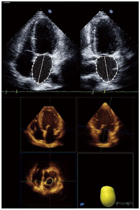
Figure 4 Left atrial volume quantification using two-dimensional (biplane Simpson’s method - upper panel) and three-dimensional echocardiography (lower panel).
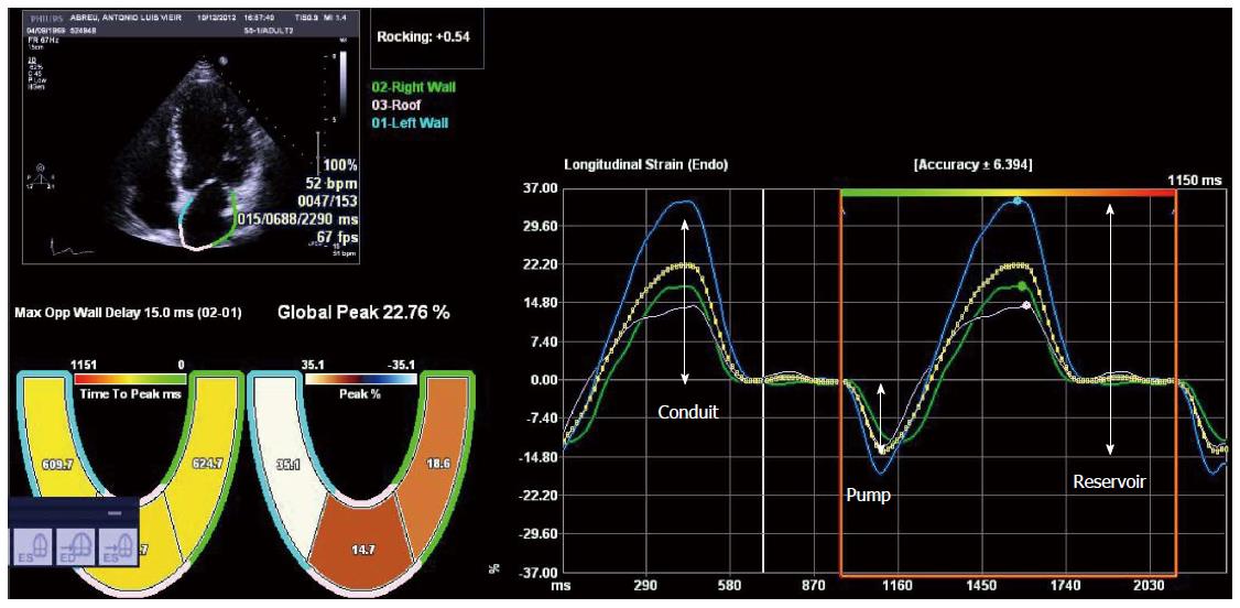
Figure 5 Left atrial deformation analysis using speckle tracking echocardiography.
Reservoir, conduit and pump function of the left atrium during the cardiac cycle can be quantified from the strain curves.
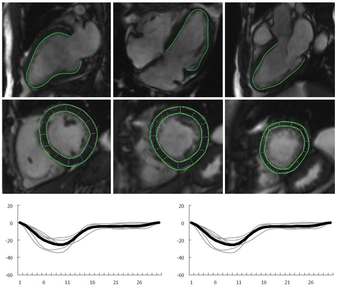
Figure 6 Left ventricular deformation analysis using magnetic resonance feature tracking.
- Citation: Sampaio F, Pimenta J. Left ventricular function assessment in cirrhosis: Current methods and future directions. World J Gastroenterol 2016; 22(1): 112-125
- URL: https://www.wjgnet.com/1007-9327/full/v22/i1/112.htm
- DOI: https://dx.doi.org/10.3748/wjg.v22.i1.112


